

Introduction
Neuroinflammation is an important biological interface linking innate immune activation, neuronal stress, synaptic dysfunction, and vulnerability to cognitive impairment (Muzio et al., 2021; Tastan and Heneka, 2024). Microglia are the resident immune cells of the central nervous system and continuously survey the brain microenvironment, contributing to tissue homeostasis, clearance of cellular debris, and synaptic remodeling (Colonna and Butovsky, 2017; Pallarés-Moratalla and Bergers, 2024). However, under persistent inflammatory stimulation, microglia can acquire a pro-inflammatory phenotype characterized by increased production of cytokines, inflammatory enzymes, and oxidative mediators (Lull and Block, 2010). If sustained, this response may contribute to neuronal injury and disruption of synaptic integrity (Wang et al., 2015).
Lipopolysaccharide (LPS), a structural component of the outer membrane of Gram-negative bacteria, is widely used to induce inflammatory activation in microglial models (Dai et al., 2015; Skrzypczak-Wiercioch and Sałat, 2022). LPS activates Toll-like receptor 4-dependent signaling and downstream inflammatory pathways, including nuclear factor-κB (NF-κB) and mitogen-activated protein kinase (MAPK) cascades. These pathways regulate the expression of inflammatory mediators such as TNF-α, IL-1β, IL-6, COX-2, and iNOS (Kang et al., 2024; Kim et al., 2023; Park et al., 2020). Although BV-2 cells do not fully recapitulate the complexity of primary microglia or the in vivo brain microenvironment, they are widely used as a practical platform for preliminary screening of candidate materials that modulate anti-inflammatory and neurotrophic biomarker responses (Henn et al., 2009; Timmerman et al., 2018).
Microglial inflammatory activation is also associated with alterations in neurotrophic and synapse-related biomarkers (Prowse and Hayley, 2021). Brain-derived neurotrophic factor (BDNF) is a representative neurotrophin involved in neuronal survival, synaptic plasticity, and memory-associated processes (Cunha et al., 2010; Kowiański et al., 2018; Miranda et al., 2019). Postsynaptic density protein 95 (PSD-95) is a major postsynaptic scaffold protein associated with excitatory synapse organization, receptor anchoring, and long-term potentiation (Ehrlich and Malinow, 2004; Xu, 2011). In the present study, the expression of Dlg4 mRNA, which encodes PSD-95, was evaluated as a synapse-related biomarker together with BDNF-related markers. Therefore, a response characterized by suppression of LPS-induced inflammatory mediator production and increased BDNF-related biomarkers and Dlg4 mRNA expression can be interpreted as a biomarker profile reflecting attenuation of inflammatory activation and improvement of neurotrophic/synapse-related marker responses in BV-2 cells.
With increasing interest in the gut–microbiota–brain axis, probiotics and postbiotic-type microbial preparations have been investigated as candidate materials capable of modulating neuroimmune responses and neurotrophic pathways (Abdel-Haq et al., 2019; Baek et al., 2024). In particular, lactic acid bacteria have attracted attention in gut–brain axis-related research because of their potential to modulate immune responses, improve the intestinal environment, and regulate host-cell responses (Cristofori et al., 2021). However, when viable bacteria are directly applied to cell culture systems, bacterial viability, proliferation, metabolite production, and changes in the culture environment may influence cellular responses. In contrast, heat-killed bacterial cells can minimize the effects associated with bacterial proliferation while retaining bacterial surface structures, cell wall components, and heat-stable bacterial components. Therefore, they can be used as postbiotic-type preparations to evaluate strain-dependent host-cell responses (Geraldo et al., 2020; Piqué et al., 2019).
Strain-level identification is important in the development of microbial-based functional materials. Even within the same species, immunomodulatory activity, cellular response induction, and physiological activity may vary depending on the strain; therefore, functional properties cannot be generalized at the genus or species level (McFarland et al., 2018). IDCC 3501 is a kimchi-derived strain with reported safety and anti-inflammatory potential, and IDCC 3201 has also been reported to have safety and anti-inflammatory potential (Chae et al., 2022; Yang et al., 2021). However, it remains unclear how these strains, when applied as heat-killed bacterial cell preparations either individually or in combination, regulate microglial inflammatory responses, BDNF-related biomarkers, and Dlg4 mRNA expression.
Therefore, this study evaluated whether heat-killed IDCC 3201, heat-killed IDCC 3501, and the IDCC 3201+ IDCC 3501 mixture modulate inflammatory mediators, BDNF-related biomarkers, and Dlg4 mRNA expression in LPS-stimulated BV-2 microglial cells. To this end, inflammatory mRNA expression, inflammatory cytokine secretion, Bdnf and Dlg4 mRNA expression, BDNF secretion, and intracellular BDNF protein expression were analyzed. In particular, this study aimed to compare the biomarker response patterns induced by each single strain and by the IDCC 3201+IDCC 3501 mixture in LPS- stimulated BV-2 cells.
Materials and Methods
The samples used in this study were heat-killed Lacticaseibacillus rhamnosus IDCC 3201 (IDCC 3201), heat-killed Lactiplantibacillus plantarum IDCC 3501 (IDCC 3501), and a 1:1 mixture of heat-killed IDCC 3201 and IDCC 3501 (IDCC 3201+IDCC 3501 mixture). IDCC 3201 and IDCC 3501 were cultured in de Man, Rogosa, and Sharpe medium (MRS; BD Difco, Franklin Lakes, NJ, USA) at 37°C for 24 h. The bacterial cultures were centrifuged at 8,000 ×g for 5 min, and the collected pellets were washed with phosphate-buffered saline (PBS; Thermo Fisher Scientific, Waltham, MA, USA) and resuspended in PBS. The bacterial suspensions were adjusted to 1 × 10⁸ CFU/mL before heat treatment. Subsequently, the bacterial suspensions were heat-treated at 100°C for 30 min to prepare heat-killed bacterial cells. To confirm bacterial inactivation, the heat-treated suspensions were plated on MRS agar and incubated at 37°C for 48 h, and no colony formation was observed. The IDCC 3201+ IDCC 3501 mixture was prepared by mixing equal volumes of each heat-killed bacterial cell suspension. For the IDCC 3201+IDCC 3501 mixture, the indicated concentration represents the total heat-killed bacterial cell concentration, with each strain contributing equally.
BV-2 mouse microglial cells were cultured in Dulbecco’s modified Eagle’s medium (DMEM; Thermo Fisher Scientific) supplemented with 10% fetal bovine serum (FBS; Thermo Fisher Scientific) and 1% penicillin/ streptomycin (P/S; Thermo Fisher Scientific). Cells were maintained at 37°C in a humidified incubator with 5% CO2. The culture medium was replaced every 2–3 days, and cells were used for experiments when they reached approximately 80% confluence.
The cytotoxicity of IDCC 3201, IDCC 3501, and the IDCC 3201+IDCC 3501 mixture was evaluated in BV-2 cells using the MTT assay. BV-2 cells were seeded in 96-well plates at a density of 1 × 10⁵ cells/well and incubated at 37°C for 24 h. The cells were then treated with IDCC 3201, IDCC 3501, or the IDCC 3201+IDCC 3501 mixture at concentrations of 105–107 cells/mL for 24 h.
After treatment, MTT solution (3-(4,5-dimethylthiazol- 2-yl)-2,5-diphenyltetrazolium bromide; Duchefa, Haarlem, Netherlands) was added to each well at a final concentration of 0.5 mg/mL, followed by incubation at 37°C for 2 h. The MTT solution was then removed, and the resulting formazan crystals were dissolved in dimethyl sulfoxide (DMSO; Sigma-Aldrich, St. Louis, MO, USA). Absorbance was measured at 570 nm using a microplate reader. Cell viability was calculated using the following equation:
BV-2 cells were seeded in 6-well plates and cultured for 24 h until they reached approximately 80% confluence. The cells were pretreated with IDCC 3201, IDCC 3501, or the IDCC 3201+IDCC 3501 mixture at concentrations of 105, 106, or 107 cells/mL for 1 h. Subsequently, lipopolysaccharide (LPS; Escherichia coli O111:B4; Sigma-Aldrich) was added at a final concentration of 1 μg/mL, and the cells were incubated for 24 h.
The experimental groups consisted of the non-LPS control, LPS control, IDCC 3201 105–107 cells/mL + LPS, IDCC 3501 105–107 cells/mL + LPS, and IDCC 3201+ IDCC 3501 105–107 cells/mL + LPS. Cell pellets were used for qPCR and western blotting analyses, and culture supernatants were used for ELISA.
Total RNA was extracted from BV-2 cell pellets using the RNeasy Kit (QIAGEN, Hilden, Germany). RNA quantity and purity were evaluated by measuring absorbance at 260 and 280 nm, and samples with an OD260/OD280 ratio of 1.8 or higher were used for cDNA synthesis. cDNA was synthesized using SuperiorScript III Reverse Transcriptase (Enzynomics, Daejeon, Republic of Korea) according to the manufacturer’s instructions.
qPCR was performed using SYBR Green Real-time PCR Master Mix (Toyobo, Osaka, Japan) on a QuantStudio 3 Real-Time PCR System (Applied Biosystems, Thermo Fisher Scientific). Relative mRNA expression levels were calculated using the 2−ΔΔCt method, and the primer sequences used in this study are listed in Table 1. The qPCR cycling conditions were as follows: 95°C for 10 min, followed by 40 cycles of 95°C for 15 s, annealing at 60°C for 20 s, and 72°C for 30 s.
Culture supernatants were collected after 1 h of sample pretreatment and 24 h of LPS stimulation. The concentrations of TNF-α, IL-1β, IL-6, and BDNF were measured using corresponding ELISA kits (R&D Systems, Minneapolis, MN, USA) according to the manufacturer’s instructions.
BDNF protein expression was evaluated by western blotting. The analyzed groups were the non-LPS control, LPS control, IDCC 3201 107 cells/mL + LPS, IDCC 3501 107 cells/mL + LPS, and IDCC 3201+IDCC 3501 107 cells/mL + LPS. Cell pellets were lysed using ice-cold RIPA buffer (Thermo Fisher Scientific), and the cell lysates were centrifuged at 13,000 ×g for 30 min at 4°C to collect the supernatants. Protein concentrations were determined using a BCA protein assay kit (Thermo Fisher Scientific).
A total of 30 μg of protein from each sample was denatured at 95°C for 5 min and separated on a 10% SDS-PAGE gel. The separated proteins were transferred onto a polyvinylidene difluoride (PVDF) membrane (Bio-Rad, Hercules, CA, USA), and the membrane was blocked with 3% bovine serum albumin (BSA; Sigma-Aldrich). The membrane was then incubated overnight at 4°C with an anti-BDNF primary antibody (1:1,000; catalog no. ab108319; Abcam, Cambridge, UK). β-actin was used as an internal loading control and detected using an anti-β-actin antibody (1:5,000; catalog no. A5441; Sigma-Aldrich).
After primary antibody incubation, the membrane was washed and incubated with an HRP-conjugated goat anti-rabbit IgG secondary antibody (1:5,000; catalog no. ab6721; Abcam) and an HRP-conjugated goat anti-mouse IgG secondary antibody (1:3,000; catalog no. ab6789; Abcam) at room temperature for 1 h. Protein bands were visualized using enhanced chemiluminescence (ECL) reagent (Bio-Rad). BDNF band intensity was quantified using ImageJ software (National Institutes of Health, Bethesda, MD, USA) and normalized to β-actin band intensity.
Data are presented as the mean ± SD from three independent experiments. Statistical analyses were performed using GraphPad Prism software (GraphPad Software, San Diego, CA, USA). One-way analysis of variance (ANOVA) followed by Dunnett’s multiple comparisons test was used to compare treatment groups with the LPS control. For the cell viability assay, treatment groups were compared with the untreated control. To directly compare the IDCC3201+IDCC3501 mixture with each single-strain treatment at the highest tested concentration (107 cells/mL), one-way ANOVA followed by Šídák’s multiple comparisons test was performed for selected pairwise comparisons. A value of p <0.05 was considered statistically significant. In the figures, statistical significance from Dunnett’s multiple comparisons test is indicated as *p <0.05, **p <0.01, and ***p <0.001 versus the LPS-treated control or untreated control, as appropriate. Statistical significance from Šídák’s multiple comparisons test is indicated as #p <0.05, ##p <0.01, and ###p <0.001 between the indicated groups.
Results
The cytotoxicity of IDCC 3201, IDCC 3501, and the IDCC 3201+IDCC 3501 mixture was evaluated in BV-2 cells using the MTT assay (Fig. 1). BV-2 cells were treated with IDCC 3201, IDCC 3501, or the IDCC 3201+IDCC 3501 mixture at concentrations of 105–107 cells/mL for 24 h. Cell viability was maintained at levels comparable to the untreated control in all treatment groups.
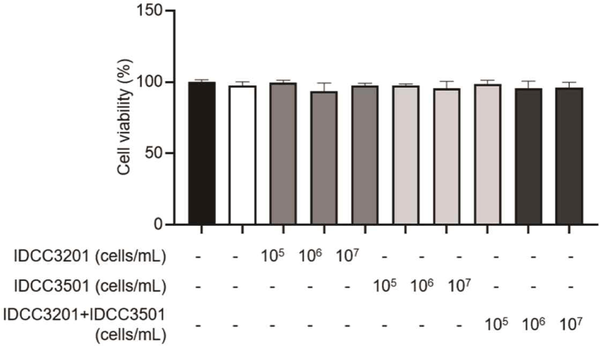
These results indicate that the test materials did not induce non-specific cytotoxicity in BV-2 cells under the present experimental conditions. Therefore, the concentration range of 105–107 cells/mL was used for subsequent experiments.
To evaluate the anti-inflammatory effects of IDCC 3201, IDCC 3501, and the IDCC 3201+IDCC 3501 mixture, the mRNA expression of inflammatory genes, including Tnf, Il1b, Il6, and Ptgs2, was measured in LPS-stimulated BV-2 cells (Fig. 2). LPS stimulation markedly increased the expression of these inflammatory genes, confirming that the inflammatory activation model was successfully established in BV-2 microglial cells.
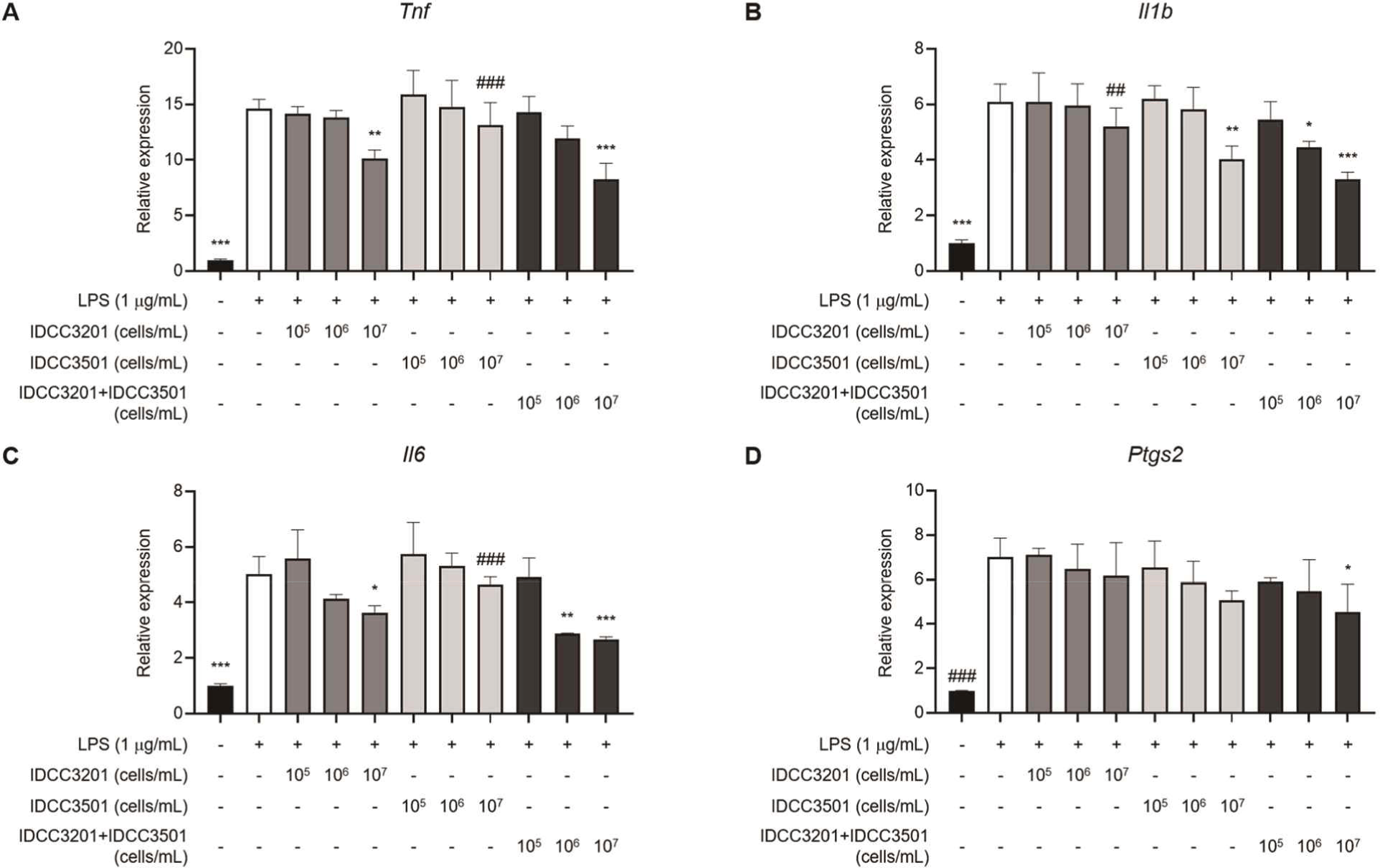
Among the treatment groups, the IDCC 3201+IDCC 3501 mixture showed the most consistent suppressive pattern across the four inflammatory genes. At 107 cells/mL, the mixture significantly reduced the mRNA expression of Tnf, Il1b, Il6, and Ptgs2 by 43.3% (p = 0.0002), 45.4% (p = 0.0001), 47.2% (p = 0.0004), and 35.0% (p = 0.0377), respectively, compared with the LPS control.
The single-strain treatments showed relatively selective responses. IDCC 3201 at 107 cells/mL significantly reduced Tnf and Il6 mRNA expression, whereas IDCC 3501 at 107 cells/mL significantly reduced Il1b mRNA expression. In contrast, the IDCC 3201+IDCC 3501 mixture significantly modulated all four inflammatory transcripts. At 107 cells/mL, direct comparisons using Šídák’s multiple comparisons test identified significant differences between the IDCC 3201+IDCC 3501 mixture and the single-strain treatments for selected inflammatory transcripts, as indicated in Fig. 2. These results indicate that the mixture modulated multiple inflammatory transcripts, while the direct differences from the single-strain treatments were marker-dependent.
The mRNA expression of the neurotrophic and synapse-related markers Bdnf and Dlg4 was evaluated in LPS-stimulated BV-2 cells (Fig. 3). LPS stimulation reduced the expression of these markers, indicating that inflammatory activation was accompanied by decreased neurotrophic and synapse-related biomarker expression in BV-2 cells. Dlg4 encodes postsynaptic density protein 95 (PSD-95) and was interpreted as a synapse-related biomarker in this study.
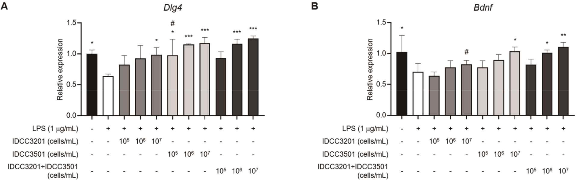
LPS treatment decreased Dlg4 mRNA expression from 1.00 ± 0.06 in the non-LPS control to 0.64 ± 0.03 in the LPS control. Bdnf mRNA expression was also decreased to 0.71 ± 0.13 in the LPS control. These results indicate that LPS induced an inflammatory response while reducing BDNF/PSD-95-related marker expression in BV-2 cells.
Bacterial treatment partially restored these LPS-induced reductions. IDCC 3201 at 107 cells/mL significantly increased Dlg4 mRNA expression by 53.9% relative to the LPS control (p = 0.0211), whereas the increase in Bdnf mRNA expression was not statistically significant. IDCC 3501 at 107 cells/mL increased Dlg4 and Bdnf mRNA expression by 82.0% (p = 0.0003) and 46.8% (p = 0.0152), respectively.
The IDCC 3201+IDCC 3501 mixture showed the most consistent restoration pattern across both markers. The mixture increased Dlg4 mRNA expression by 94.1% (p <0.0001) and Bdnf mRNA expression by 56.4% (p = 0.0029) relative to the LPS control. These findings indicate that the IDCC 3201+IDCC 3501 mixture partially restored LPS-suppressed Bdnf and Dlg4 mRNA expression. Direct comparisons at 107 cells/mL identified significant differences between the mixture and the single-strain treatments for selected markers, as indicated in Fig. 3.
To determine whether the suppression of inflammatory mRNA expression was reflected at the cytokine secretion level, the concentrations of TNF-α, IL-1β, and IL-6 in culture supernatants were measured by ELISA (Fig. 4). LPS stimulation markedly increased the secretion of all three inflammatory cytokines.
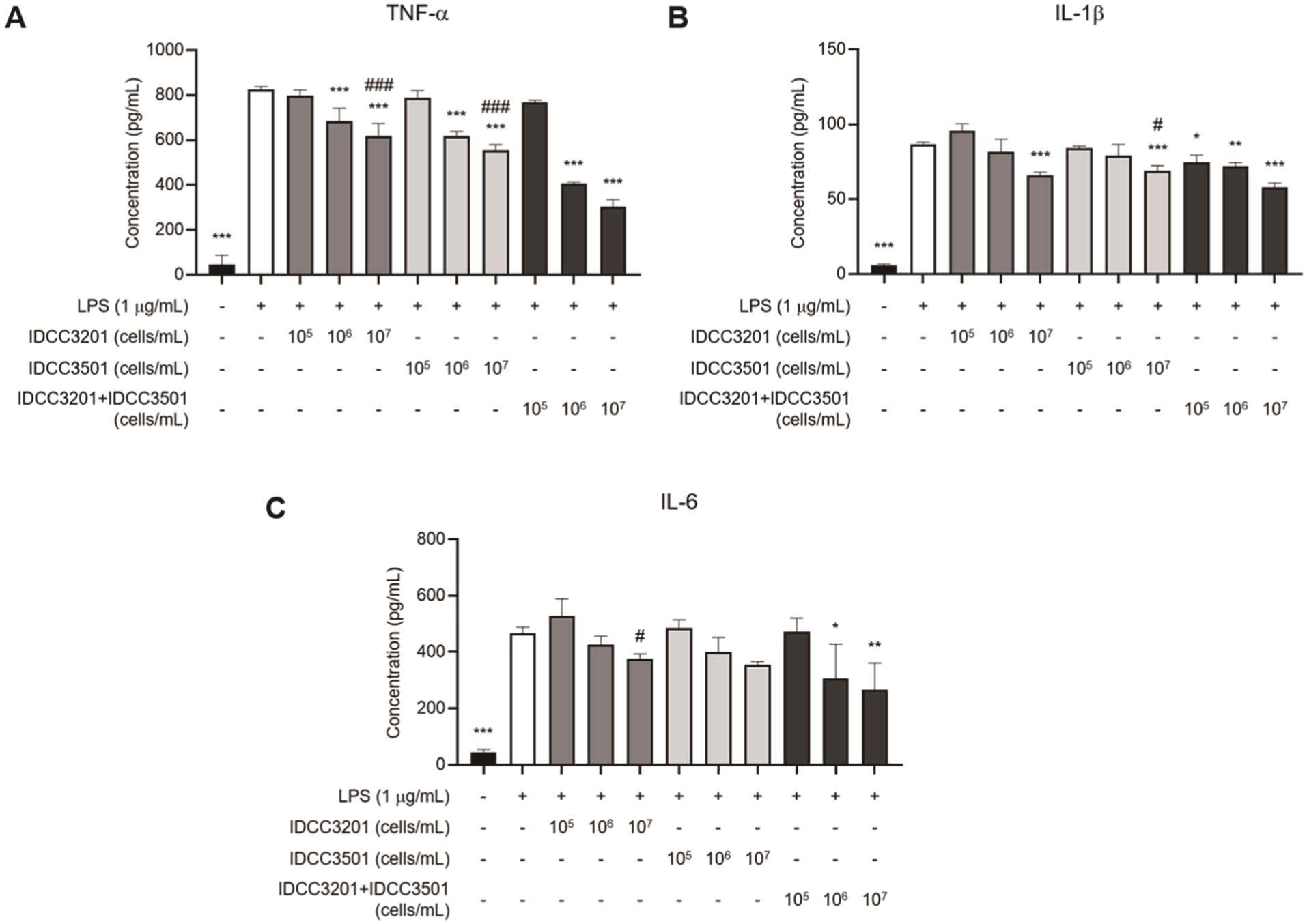
TNF-α secretion increased from 47.10 ± 40.96 pg/mL in the non-LPS control to 823.81 ± 13.65 pg/mL in the LPS control. IDCC 3201 and IDCC 3501 at 107 cells/mL reduced TNF-α secretion to 616.05 ± 57.88 pg/mL and 553.62 ± 25.58 pg/mL, respectively. The mixture reduced TNF-α secretion to 303.81 ± 32.60 pg/mL, corresponding to a 63.1% decrease relative to the LPS control (p <0.0001).
IL-1β secretion increased from 5.63 ± 1.20 pg/mL in the non-LPS control to 86.48 ± 1.62 pg/mL in the LPS control. IDCC 3201 and IDCC 3501 at 107 cells/mL reduced IL-1β secretion to 65.93 ± 2.10 pg/mL and 69.02 ± 3.28 pg/mL, respectively. The mixture reduced IL-1β secretion to 57.86 ± 3.10 pg/mL, corresponding to a 33.1% decrease relative to the LPS control (p <0.0001).
IL-6 secretion increased from 43.70 ± 12.23 pg/mL in the non-LPS control to 468.30 ± 20.86 pg/mL in the LPS control. Although IDCC 3201 and IDCC 3501 reduced the mean IL-6 level at 107 cells/mL, these reductions did not reach statistical significance. In contrast, the mixture significantly reduced IL-6 secretion to 265.67 ± 95.17 pg/mL, corresponding to a 43.3% decrease relative to the LPS control (p = 0.0019). At 107 cells/mL, direct comparisons showed significant differences between the mixture and the single-strain treatments for selected cytokines, as indicated in Fig. 4.
These results indicate that the IDCC 3201+IDCC 3501 mixture suppressed not only LPS-induced inflammatory mRNA expression but also extracellular inflammatory cytokine release.
To further evaluate the neurotrophic biomarker response to bacterial treatment, BDNF levels in culture supernatants were measured by ELISA, and intracellular BDNF protein expression was evaluated by western blotting. LPS stimulation reduced BDNF secretion from 590.20 ± 17.81 pg/mL in the non-LPS control to 421.70 ± 57.87 pg/mL in the LPS control, indicating that inflammatory activation decreased the BDNF-related neurotrophic biomarker response in BV-2 cells (Fig. 5A).
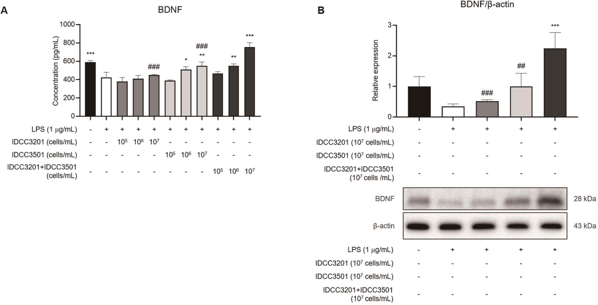
Among the single-strain treatments, IDCC 3501 at 107 cells/mL increased secreted BDNF to 548.98 ± 43.53 pg/mL, whereas IDCC 3201 did not significantly increase BDNF secretion. The IDCC 3201+IDCC 3501 mixture increased BDNF secretion to 753.60 ± 48.27 pg/mL, corresponding to a 78.7% increase relative to the LPS control (p <0.0001).
Western blotting also supported the BDNF-restoring response of the mixture (Fig. 5B). LPS reduced intracellular BDNF protein expression from 1.00 ± 0.32 in the non-LPS control to 0.35 ± 0.08 in the LPS control. IDCC 3201 increased the mean BDNF protein expression level to 0.52 ± 0.05, and IDCC 3501 increased it to 1.00 ± 0.43; however, neither single-strain treatment reached statistical significance. In contrast, the mixture increased intracellular BDNF protein expression to 2.24 ± 0.52, corresponding to an approximately 6.4-fold increase relative to the LPS control (p = 0.0002).
Taken together, the IDCC 3201+IDCC 3501 mixture suppressed LPS-induced inflammatory cytokine responses while increasing both BDNF secretion and intracellular BDNF protein expression. Direct comparisons at 107 cells/mL further identified significant differences between the IDCC 3201+IDCC 3501 mixture and the single-strain treatments for BDNF-related measurements, as indicated in Fig. 5. Taken together, the mixture was associated with reduced inflammatory cytokine responses and increased BDNF-related biomarker responses in LPS-stimulated BV-2 cells.
The integrated profiles summarize changes in (A) inflammatory biomarkers, including Tnf, Il1b, Il6, and Ptgs2 mRNA expression and TNF-α, IL-1β, and IL-6 secretion, and (B) BDNF/Dlg4-related biomarkers, including Dlg4 and Bdnf mRNA expression, BDNF secretion, and intracellular BDNF protein expression. Biomarker changes at 107 cells/mL were visualized relative to the LPS-treated control.
The integrated biomarker profile is presented in Fig. 6. Fig. 6A shows the inflammatory biomarker profile, including the inflammatory transcripts Tnf, Il1b, Il6, and Ptgs2, as well as the secreted cytokines TNF-α, IL-1β, and IL-6. Fig. 6B summarizes the BDNF- and Dlg4-related profile, including Bdnf mRNA expression, BDNF secretion, intracellular BDNF protein expression, and Dlg4 mRNA expression.
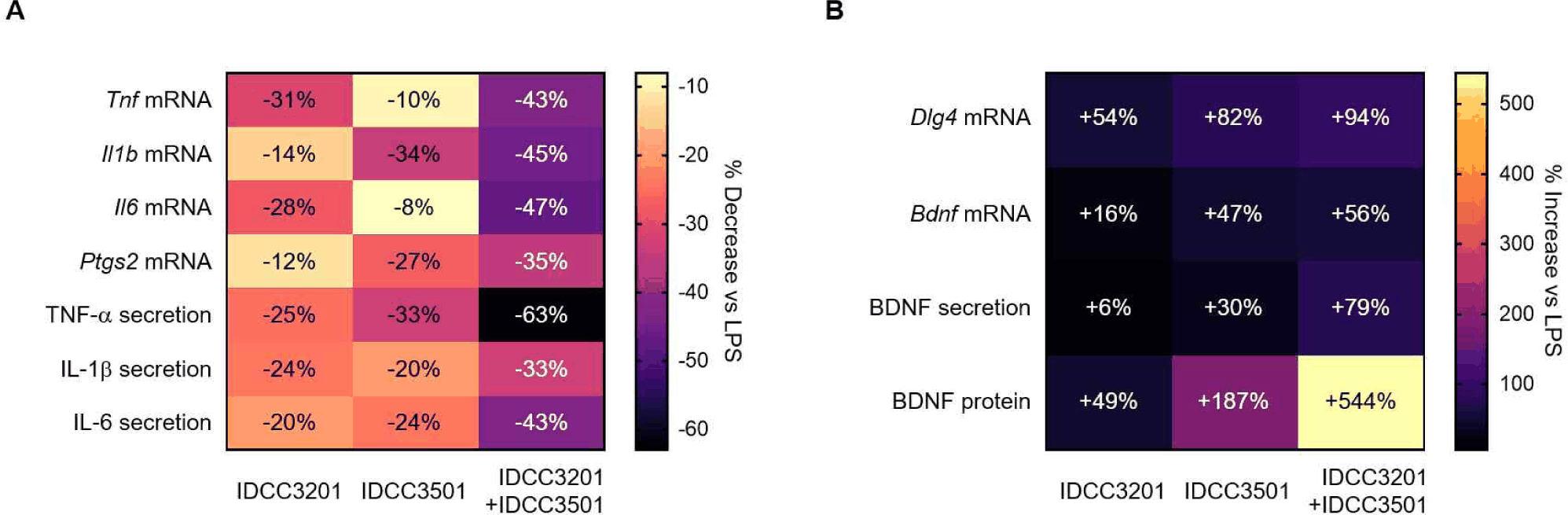
Compared with the LPS control, the IDCC 3201+IDCC 3501 mixture showed an overall reduction in inflammatory markers. Specifically, the mixture reduced the mRNA expression of Tnf, Il1b, Il6, and Ptgs2, together with the secretion of TNF-α, IL-1β, and IL-6. In contrast, BDNF- and Dlg4-related markers showed an increasing pattern, indicating that the mixture restored LPS-suppressed BDNF-related biomarkers and Dlg4 mRNA expression.
In the integrated profile, IDCC 3201 showed a relatively selective reduction in inflammatory markers, whereas IDCC 3501 showed a more evident increase in BDNF- and Dlg4-related markers. The IDCC 3201+IDCC 3501 mixture combined these partial response patterns into a broader biomarker profile characterized by reduced inflammatory markers and increased BDNF/Dlg4-related markers. These results suggest that the IDCC 3201+IDCC 3501 mixture may shift LPS-stimulated BV-2 cells toward a less inflammatory and more BDNF-related biomarker state.
Discussion
In this study, we evaluated whether heat-killed IDCC 3201, heat-killed IDCC 3501, and the IDCC 3201+IDCC 3501 mixture modulate inflammatory mediators, BDNF-related biomarkers, and Dlg4 mRNA expression in LPS-stimulated BV-2 microglial cells. The major finding of this study was that the IDCC 3201+IDCC 3501 mixture reduced the expression of inflammation-related mRNAs and the secretion of inflammatory cytokines, while increasing BDNF secretion, intracellular BDNF protein expression, and Dlg4 mRNA expression. These results suggest that the heat-killed IDCC 3201+IDCC 3501 mixture may attenuate the LPS-induced inflammatory biomarker profile and promote the recovery of BDNF-related neurotrophic marker responses.
LPS induces a pro-inflammatory response in microglia through TLR4-dependent signaling and increases the expression of inflammatory mediators such as TNF-α, IL-1β, IL-6, and COX-2 (Fang et al., 2022; Li et al., 2022; Skrzypczak-Wiercioch and Sałat, 2022). In the present study, LPS stimulation increased the mRNA expression of Tnf, Il1b, Il6, and Ptgs2, as well as the secretion of TNF-α, IL-1β, and IL-6. In contrast, the IDCC 3201+IDCC 3501 mixture broadly reduced these inflammation-related transcripts and cytokines. The fact that the mixture showed a consistent inhibitory pattern across a wider range of markers than either single strain suggests that its effect was not limited to a specific cytokine, but rather reflected broader modulation of the LPS-induced inflammatory biomarker profile.
The differences in responses between the single strains and the mixture are also noteworthy. IDCC 3201 showed relatively pronounced inhibitory effects on several inflammation-related markers, whereas IDCC 3501 showed relatively clearer effects on BDNF- and Dlg4-related markers. The IDCC 3201+IDCC 3501 mixture showed a broader biomarker-modulating profile across inflammatory and BDNF/Dlg4-related markers, suggesting that the two strains may contribute differently to the overall response pattern. Previous studies have discussed that multi-strain probiotic or postbiotic preparations may provide broader biological effects through combined or complementary strain-specific properties (Chapman et al., 2011; Kwoji et al., 2021). In the present study, additional direct comparisons at 107 cells/mL identified significant differences between the mixture and the single-strain treatments for selected biomarkers. However, these selected pairwise comparisons were limited to the highest tested concentration and do not establish the overall statistical superiority or synergistic activity of the mixture. Therefore, the mixture response should be interpreted as a broad and coordinated biomarker-modulating profile rather than definitive evidence of synergy.
The recovery of BDNF-related markers is an important feature of this study. BDNF is a representative neurotrophin associated with neuronal survival, synaptic plasticity, and memory-associated processes. In this study, LPS reduced Bdnf mRNA expression, BDNF secretion, and intracellular BDNF protein expression, whereas the IDCC 3201+IDCC 3501 mixture increased these markers at multiple levels. In particular, intracellular BDNF protein expression was significantly increased only in the mixture-treated group, supporting the possibility that the mixture more effectively restored BDNF-related biomarker responses. These findings suggest that, under inflammatory microglial activation, the mixture may be involved not only in the suppression of inflammatory responses but also in the recovery of the neurotrophic biomarker profile.
The changes in Dlg4 mRNA expression further suggest that synapse-related biomarkers may be affected under LPS-induced inflammatory conditions (Badshah et al., 2016; Sheppard et al., 2019). Dlg4 encodes PSD-95, a postsynaptic scaffold protein involved in excitatory synapse organization and postsynaptic scaffolding (Levy et al., 2022). In this study, LPS decreased Dlg4 mRNA expression, whereas the IDCC 3201+IDCC 3501 mixture increased its expression. However, because PSD-95 protein expression and synaptic function were not directly evaluated, this result should be interpreted at the level of a synapse-related biomarker.
The samples used in this study were heat-killed bacterial cells prepared by treatment at 100°C for 30 min. Therefore, the observed effects are likely to have been mediated by bacterial surface structures, cell wall components, or heat-stable bacterial components rather than by bacterial proliferation or newly produced metabolites during treatment (Bron et al., 2013; Piqué et al., 2019). Unlike viable bacteria, heat-killed bacterial cells can reduce confounding factors associated with bacterial growth, acid production, and changes in the culture medium composition. Thus, they are useful for evaluating host-cell responses induced by bacterial cellular components in cell-based models (Geraldo et al., 2020; Piqué et al., 2019). In addition, postbiotic-type preparations may have advantages over viable bacteria in terms of stability, safety, and applicability, which is meaningful from the perspective of functional material development (Aguilar-Toalá et al., 2018). In this context, the heat-killed IDCC 3201+IDCC 3501 mixture can be considered a postbiotic-type candidate material capable of modulating inflammatory and BDNF-related biomarker responses in BV-2 cells.
Several limitations should be considered when interpreting the findings of this study. First, BV-2 cells are an immortalized microglial cell line and do not fully represent primary microglia or the complexity of the in vivo brain microenvironment (Henn et al., 2009; Stansley et al., 2012). Second, although this study evaluated changes in inflammatory mediators and BDNF/Dlg4-related biomarkers, it did not directly assess neuronal protection, synaptic function, or cognitive function. Third, the upstream mechanisms underlying the observed changes remain unclear because related signaling pathways, such as NF-κB/MAPK and TrkB/CREB/Akt, were not directly analyzed. Therefore, the present findings should be interpreted as phenotypic modulation of inflammatory and BDNF/ Dlg4-related biomarkers rather than direct evidence of pathway-level regulation. Fourth, PSD-95 was evaluated only at the Dlg4 mRNA level, not at the protein level, and the specific active components of the heat-killed bacterial cells responsible for the observed effects were not identified. Fifth, this study did not include a known anti-inflammatory or microglial-modulating positive control, which limits the ability to benchmark the magnitude of the observed effects against a reference treatment. Future studies should investigate inflammatory signaling, BDNF-related signaling, PSD-95 protein expression, and functional validation using neuron–microglia interaction models.
In conclusion, this study demonstrates that the IDCC 3201+IDCC 3501 mixture reduced inflammatory transcripts and cytokine secretion while increasing BDNF-related biomarkers and Dlg4 mRNA expression in LPS-stimulated BV-2 microglial cells. The mixture showed a broad and coordinated biomarker-modulating pattern across inflammatory and BDNF-related markers, suggesting its potential as a postbiotic-type candidate material for further investigation. However, these findings provide preliminary evidence at the in vitro biomarker level, and further pathway-level studies and in vivo validation are required before linking these biomarker responses to actual neuroprotective or cognitive efficacy.


