Current Topic in Lactic Acid Bacteria and Probiotics
Korean Society for Lactic Acid Bacteria and Probiotics
Research Article

김치 유래 Lactiplantibacillus plantarum 사균체의 항산화 및 항염 효과

유청빈1https://orcid.org/0000-0002-5301-7100, 이은수1https://orcid.org/0000-0003-4613-8999, 이민경1https://orcid.org/0000-0002-4802-5739, 이가영1https://orcid.org/0000-0002-7050-7101, 박훈1,*https://orcid.org/0000-0002-8965-8196
Cheong-Bin You1https://orcid.org/0000-0002-5301-7100, Eun-Su Lee1https://orcid.org/0000-0003-4613-8999, Min-Kyeong Lee1https://orcid.org/0000-0002-4802-5739, Ga-Young Lee1https://orcid.org/0000-0002-7050-7101, Hoon Park1,*https://orcid.org/0000-0002-8965-8196
1선문대학교 식품과학과
1Department of Food Science, Sun Moon University, Asan 31460, Republic of Korea
*Corresponding author : Hoon Park, Department of Food Science, Sun Moon University, Asan 31460, Korea. Tel: +82-41-530-2262, Fax: +82-41-530-2917, E-mail: hpark@sunmoon.ac.kr

Copyright © Korean Society for Lactic Acid Bacteria and Probiotics. This is an Open-Access article distributed under the terms of the Creative Commons Attribution Non-Commercial License (http://creativecommons.org/licenses/by-nc/4.0/) which permits unrestricted non-commercial use, distribution, and reproduction in any medium, provided the original work is properly cited.

Received: Nov 01, 2022; Revised: Dec 01, 2022; Accepted: Dec 06, 2022

Published Online: Dec 31, 2022

Abstract

The aim of this study was to evaluate the probiotic properties of lactic acid bacteria (LAB) isolated from kimchi and antioxidant and anti-inflammatory activities of heat-killed LAB strains. Regarding probiotic properties, Lactiplantibacillus plantarum SMF398 and L. plantarum SMF470 isolated from kimchi can survive under artificial gastric condition and adhere strongly to HT-29 cells. The SMF398 and SMF470 strains showed strong antimicrobial activity and co-aggregation against pathogenic bacteria. The heat-killed cells of LAB (1 mg/mL) were prepared by heating at 121°C for 15 min. MTT assay revealed that the heat-killed cells (1 mg/mL) of SMF398 and SMF470 were not toxic to HT-29 cells. The heat-killed SMF398 and SMF470 showed significantly higher DPPH and ABTS radical scavenging activities as well as β-carotene bleaching inhibitory activity than the heat-killed L. plantarum ATCC14917, a control probiotic strain (p<0.05). In lipopolysaccharide-induced RAW264.7 cells, the heat-killed SMF398 and SMF470 significantly reduced the nitric oxide production by 30.92% and 22.81%, respectively (p<0.05). Furthermore, the heat-killed SMF398 and SMF470 significantly decreased the gene expression levels of tumor necrosis factor-α, interleukin-6, inducible nitric oxide synthase, cyclooxygenase-2 up to 58.02%, 67.70%, 53.44%, 45.90%, respectively (p<0.05). These results suggest that the heat-killed L. plantarum SMF398 and L. plantarum SMF470 might be useful as antioxidant and anti-inflammatory agents.

Keywords: Lactiplantibacillus plantarum; paraprobiotics; heat-killed probiotics; antioxidant activity; anti-inflammatory activity

서 론

프로바이오틱스(probiotics)는 숙주의 건강에 도움을 주는 살아있는 미생물로, 기본적으로 유해균 억제, 유익균 증식 및 배변활동에 도움을 주며, 균주의 특성에 따라 면역과민반응 조절, 여성 질 건강 개선, 체지방 감소, 콜레스테롤 감소, 중추신경계 기능 향상 등 다양한 생리활성을 나타낸다(Choi, 2019; Moon, 2019). 프로바이오틱스로 Lactobacillus 속, Bifidobacterium 속, Enterococcus 속 등의 미생물이 대표적으로 이용되고 있다. 프로바이오틱스 균주로 이용되기 위해서는 비병원성이고 돌연변이 유발이나 용혈능이 없어야 하며 항생제에 대한 내성이 없는 등 안전성이 확보되어야 한다(Chu and Kook, 2019). 또한 인간의 장내에서 생리활성을 발휘하기 위해서는 위산 및 담즙에 대한 생존력이 높아야 하고, 장 상피세포에 부착력이 강해야 하며, 병원성 미생물을 억제할 수 있는 항균 물질 등을 생산해야 한다(Almudena et al., 2014). 세계적으로 프로바이오틱스 시장이 급성장하고 있으나, 생균의 사용에 대한 오남용으로 설사, 변비, 복통, 복부팽만감, 구토, 피부 홍반 및 두드러기와 같은 부작용 사례가 지속적으로 증가하고 있고, 중증의 사례로 일부 고위험군에서 패혈증과 균혈증이 보고되었다(Kim et al., 2020a). 이러한 이유로 생균보다 안전하며 생균제 사용이 어려운 장누수증 환자 또는 기저질환 환자에게 대체제로 사용 가능한 프로바이오틱스의 사균체(파라프로바이오틱스, paraprobiotics)와 대사산물(포스트바이오틱스, postbiotics) 연구가 전 세계적으로 관심을 받고 있다.

파라프로바이오틱스(paraprobiotics)는 물리적 또는 화학적 방법에 의해 비활성화된 기능성 미생물의 사균체를 말한다. 사균체는 생균에 비하여 안전성(safety)과 안정성(stability)이 높으며, 천연첨가물로써 식품, 제약, 화장품 등 다양한 분야에 적용할 수 있는 장점이 있다. 유산균 사균체 제조 방법으로 열처리, 자외선, 고압, 초음파, 고전압 펄스 전기장(pulsed electric field), 옴가열 기술(ohmic heating process) 등이 연구되었으며, 그 중에서 열처리 방법은 공정이 단순하고 미생물의 완전한 멸균이 가능하며 온도처리 조건에 따라 세포벽의 파괴 정도를 조절할 수 있는 장점이 있어 가장 보편적으로 이용되고 있다(Barros et al., 2021; Ou et al., 2011; Piqué et al., 2019). 최근의 연구에 의하면 프로바이오틱스의 열처리 과정에서 peptidoglycans, nucleic acid, lipoteichoic acid, exopolysaccharides(EPS), cell wall protein 등 다양한 생리활성 성분이 균체로부터 용출되어 항산화 작용, 항염 작용, 장내 균총 개선, 콜레스테롤 개선, 면역 조절, 인지능력 향상 등 생균에 준하는 효능을 나타낸다고 보고되었다(Akter et al., 2020; Nataraj et al., 2020; Piqué et al., 2019). Lactobacillus 속 균주에서 추출한 peptidoglycan이 항염과 면역조절 작용을 보였으며(Shida et al., 2009), EPS는 열에 대한 안정성이 높고 항산화, 항염, 면역조절 작용 등 다양한 효능을 나타내었다고 보고되었다(Lee et al., 2015; Son and Kang, 2021; Yang and Kim, 2001). Lactobacillus paracasei D3-5의 열처리 사균체가 대사증후군이 유발된 고령의 쥐에서 장내 염증을 완화하고 운동능력과 인지능력을 향상하였으며, 사균체에서 분리한 lipoteichoic acid가 mucin 생산 증가, 장누수 억제, 염증 완화 작용에 관여하는 것으로 보고되었다(Wang et al., 2019).

인체의 산소 소비 과정에서 불가피하게 발생하는 활성산소종(reactive oxygen species; ROS)이 산화적인 스트레스에 의해 체내에 과도하게 축적되면 세포의 구성 성분인 단백질, 지방, DNA 등을 비가역적으로 손상하여 염증을 유발하고, 암, 간경변증, 관절염, 심혈관질환, 뇌졸중, 알츠하이머 등과 같은 각종 질병을 일으킬 수 있다(Alberto et al., 2012; Reddy et al., 2009). 최근 Lactipantibacillus plantarum 열처리 사균체가 생균과 비교하여 높은 항산화활성을 보였으며(Song et al., 2019), Enterococcus faecalis 사균체를 첨가한 발효유에서 항염 활성이 농도 의존적으로 증가되었다고 보고되었다(Choi et al., 2018). 현재까지 프로바이오틱스로 이용되는 LactobacillusBifidobacterium 등의 생균이 항산화 및 항염 활성을 나타낸다는 다수의 연구 결과들이 보고되었지만(Jang et al., 2018, Kim et al., 2020b), 유산균 사균체를 활용한 국내의 생리활성 연구는 외국과 비교하여 매우 부족한 실정이다. 따라서 김치, 젓갈 등 전통발효식품에서 분리된 유산균의 열처리 사균체를 활용하여 광범위한 생리활성 연구가 필요하다.

본 연구에서는 김치로부터 분리된 유산균 중 내산성, 내담즙성, 항균활성 및 장부착능이 우수한 2종의 균주를 선별하고 프로바이오틱스 활성을 평가하였다. 또한 선별균주를 열처리하여 사균체의 항산화 활성과 항염 활성을 측정함으로써 활성산소와 염증에 의해 유발되는 각종 질병들을 예방하기 위한 기능성 사균체 개발의 기초자료로 활용하고자 하였다.

재료 및 방법

실험재료

본 연구에서 사용된 김치는 천안과 아산의 전통시장에서 구매하여 사용하였다. 대조군으로 사용된 프로바이오틱스 유산균주인 Lactiplatibacillus plantarum ATCC14917(KCTC3108)과 병원성 지시세균인 Escherichia coli KCTC1039는 생물자원센터(Daejeon, Korea)에서, Staphylococcus aureus KCCM11335는 한국미생물보존센터(Seoul, Korea)에서 분양받았다. 인간 결장 유래 HT-29 세포(KCLB30038)와 마우스 유래 대식세포인 RAW264.7 세포(KCLB40071)는 한국세포주은행(Seoul, Korea)에서 분양받았다. DPPH(1,1-diphenyl1-2-picrylhydrazyl), ABTS(2,2'-azino-bis 3-ethylbenzothiazoline-6-sulphonic acid), MTT(3-(4,5-dimethyl thiazol-2-yl)-2,5-diphenyl tetrazolium bromide), LPS(lipopolysaccharide), pepsin, oxgall, sulfanilamide, phosphoric acid, naphthylethylene diamine dihydrochloride 등의 시약은 Sigma-Aldrich Co.(Saint Louis, USA)에서 구입하였다.

유산균의 분리 및 동정

김치시료를 멸균 생리식염수에 1:9(w/v) 비율로 희석한 후 Stomacher(BagMixer 400W, Interscience, Saint Nom, France)를 사용하여 5분간 마쇄하였다. 마쇄한 용액을 십진법으로 희석한 후 DeMan Rogosa Sharpe(MRS, DifcoTM, MD, USA) 고체배지에 도말하고 37°C에서 overnight 배양하였다. 배양 후 크기가 큰 단일 콜로니를 BCP 고체배지(Eiken chemical Co., Ltd., Tokyo, Japan)에 접종하여 배양한 후 젖산 생성으로 인해 노란색 환을 나타내는 콜로니를 유산균으로 간주하고 선별하였다. 현미경(AE31 Trinocular, Microscope World, Carlsbad, USA)을 이용하여 선별된 유산균주의 형태학적 특성을 관찰하고, Gram 염색성과 catalase 활성을 확인하였다. 선별된 유산균주의 16S rRNA 염기서열 분석은 BIOFACT사(Daejeon, Korea)를 통하여 진행하였다. 유전자 증폭은 universal rRNA gene primer(27F and 1492R)를 이용하였고 전기영동을 통하여 PCR 산물의 증폭 여부를 확인하였다. 16S rRNA 염기서열 분석 결과는 National Center for Biotechnology Institute(NCBI)의 BLAST online program을 이용하여 Genbank database와 비교하였다. 선별된 유산균은 MRS 액체배지에 배양한 후 30%(v/v) glycerol stock으로 –80°C의 deep freezer(NF-140SF, Nihon Freezer Co., LTD., Tokyo, Japan)에 보관하여 사용하였다.

세포 배양

HT-29 세포는 Roswell Park Memorial Institue 1640(RPMI, Welgene, Korea)에 10% Fetal Bovine Serum(FBS, GW Vitek, Seoul, Korea)과 1% penicillin-streptomycin solution (Cytiva, Marlborough, USA)을 첨가하여 배양하였다. RAW264.7 세포는 Dulbecco’s modified Eagle’s medium(DMEM, Welgene, Gyeongsan, Korea)에 10% FBS와 1% penicillin-streptomycin solution을 첨가하여 배양하였다. 두 세포주는 37°C와 5% CO2를 유지하는 incubator(Thermo Fisher, Massachusetts, USA)에서 배양하였다.

유산균의 내산성 및 내담즙성 측정

유산균의 내산성 및 내담즙성은 Lertcanawanichakul 등(2015)의 방법을 일부 수정하여 측정하였다. MRS 액체배지에 배양(37°C, overnight)한 유산균을 약 1×10⁹ CFU/mL 수준으로 조정하여 실험에 사용하였다. 내산성 평가는 0.3% pepsin이 첨가된 MRS 액체배지(pH 2.5)에 유산균 배양액을 약 1×10⁷ CFU/mL 수준이 되도록 1%(v/v) 접종하여 3시간 반응시키고, MRS 고체배지에 도말한 후 생균수를 측정하여 생존율을 확인하였다. 내담즙성 평가는 0.3% oxgall(DifcoTM, MD, USA)을 첨가한 MRS 액체배지에 유산균 배양액을 1×10⁷ CFU/mL 수준이 되도록 1%(v/v) 접종하여 24시간 반응시키고, MRS 고체배지에 도말한 후 생균수를 측정하여 생존율을 확인하였다.

Survival rate ( % ) = ( Viable cell / Initial cell ) × 100
유산균의 항균활성 측정

유산균의 항균활성은 paper disc법으로 측정하였다(Kim et al., 1999). MRS 액체배지에서 37°C, overnight 배양한 유산균을 원심분리(12,000 ×g, 4°C, 10 min, CT15RE, Hitachi Chemical Co., Ltd., Tokyo, Japan)하여 균체를 제거한 후 0.45 μm syringe filter로 여과한 상등액을 실험에 사용하였다. 병원성 지시세균은 tryptic soy broth(TSB, DifcoTM, MD, USA)에서 37°C, overnight 배양한 후 0.85% saline으로 1×10⁶ CFU/mL 수준까지 희석한 다음 멸균된 면봉을 이용하여 nutrient agar(NA. DifcoTM, MD, USA)에 도말하였다. 병원성 지시균주가 도말된 평판배지에 8 mm 직경의 paper disc를 올려놓고 유산균 상등액을 100 μL씩 접종한 후 37°C에서 overnight 배양하였다. 유산균의 항균활성은 paper disc 주변에 형성된 생육 억제환의 크기를 측정하여 평가하였다.

유산균과 병원성 세균의 co-aggregation 측정

유산균의 병원성 지시균에 대한 co-aggregation은 Tareb 등(2013)의 방법을 일부 수정하여 진행하였다. MRS 액체배지에서 배양(37°C, overnight)한 유산균과 TSB에서 배양(37°C, overnight)한 병원성 지시세균(E. coli KCTC1039, S. aureus KCCM11335)을 PBS 용액(pH 7.2)으로 2번 세척하여 균체를 회수한 후 OD600 = 0.3으로 조정한 현탁액을 사용하였다. 유산균 현탁액(OD600 = 0.3)과 지시세균 현탁액(OD600 = 0.3)을 각 2 mL씩 혼합하고, 37°C에서 24시간 반응시킨 다음 600nm에서 흡광도를 측정한 후 다음의 계산식에 따라 유산균의 co-aggregation을 산출하였다.

Co aggregation ( % ) = [ 1 OD time 1 ) OD time 2 ) + OD pathogenes 3 ) 2 ] × 100
  • 1) ODtime: Optical density of mixture after 24 h

  • 2) ODLAB: Initial optical density of LAB

  • 3) ODpathogenes: Initial optical density of pathogenes

유산균의 장세포 부착능 측정

유산균의 HT-29 세포에 대한 장 부착능은 Shin 등(2015)의 방법을 일부 수정하여 진행하였다. MRS 액체배지에서 37°C, overnight 배양한 유산균을 PBS 용액(pH 7.2)으로 두 번 세척한 후 1×10⁹ CFU/mL로 농도를 조정한 현탁액을 사용하였다. 6-well plate (SPL Life Sciences Co., Ltd, Pocheon, Korea)에 HT-29 세포를 1×10⁶ cell/well 수준으로 접종하고, 24시간 배양(37°C, 5% CO2)하였다. 배양 후 상등액을 제거하고 HT-29 세포에 RPMI 1 mL와 유산균 현탁액 1 mL를 첨가하고 1시간 동안 반응하였다. 반응 후 상등액을 제거한 다음 PBS 용액(pH 7.2)을 사용하여 세포를 3회 세척하고, 200 μL의 0.1% Triton X-100(SAMCHUN, Seoul, Korea)을 처리하여 세포에 부착된 균을 회수한 후 MRS 고체배지에 도말하여 생균수를 측정하였다. 유산균의 장부착능(%)은 다음의 계산식에 따라서 산출하였다.

Cell adhesion ( % ) = ( Viable cell count 1 ) / Initial cell count 2 ) ) × 100
  • 1) Viable cell count : Number of adhesion cell count

  • 2) Initial cell count : Number of initial cell count

열처리 사균체 제조 방법

유산균의 사균 처리는 Cheon 등(2020)의 방법을 일부 수정하여 진행하였다. MRS 액체배지에 37°C, overnight 배양한 유산균 배양액을 동결건조(FDU-12000, EYELA, Korea)한 후 1 mg/mL(약 1×10⁹ CFU/mL)의 농도로 PBS 용액(pH 7.2)에 현탁하고, 121°C에서 15분간 고압 멸균하여 실험에 이용하였다.

사균체의 세포독성 측정

열처리 사균체의 세포독성 평가는 HT-29 세포를 이용하여 MTT 방법에 의해 수행하였다(Chuah et al., 2019). HT-29 세포를 96-well plate에 1×10⁵ cell/well 수준으로 접종한 후 37°C, 5% CO2에서 24시간 배양하였다. 배양 후 상등액을 제거하고 RPMI 배지 180 μL와 유산균 사균체 20 μL를 첨가하여 24시간 배양하였다. 상등액을 제거하고 PBS 용액으로 well에 남아있는 균체를 세척한 후 RPMI 배지 180 μL와 MTT 시약(5 mg/mL) 20 μL를 첨가하고 4시간 반응하였다. 상등액을 제거한 후 100 μL의 dimethyl sulfoxide(DMSO, DAEJUNG, Siheung, Korea)를 가하여 생성된 formazan 침전물을 용해시킨 다음 microplate reader(Biochrom, Cambridge, USA)를 이용하여 570 nm에서 흡광도를 측정하고, 다음의 계산식에 따라서 세포 생존율(%)을 계산하였다. 공시험은 PBS 용액(pH 7.2)을 사용하였다.

Cytotoxicity of heat-killed cell ( % ) = ( OD sample 1 ) + OD control 2 ) ) × 100
  • 1) ODsample : Optical density of sample

  • 2) ODcontrol : Optical density of control

DPPH 라디칼 소거능 측정

DPPH 라디칼 소거능은 Wang 등(2016)의 방법을 일부 수정하여 측정하였다. 시료 2 mL를 OD517=1.2로 조정한 0.4 mM DPPH 용액 2 mL와 혼합하고 실온의 암실에서 30분간 반응하였다. 반응액을 원심분리(12,000 ×g, 4°C, 10 min)하여 상등액을 회수한 후 517 nm에서 흡광도를 측정하고, 다음의 계산식에 따라서 DPPH 라디칼 소거능(%)을 계산하였다. 공시험은 PBS 용액(pH 7.2)을 사용하였다.

DPPH radical scavenging activity ( % ) = { 1 ( OD sample 1 ) + OD control 2 ) ) } × 100
  • 1) ODsample : Optical density of sample

  • 2) ODcontrol : Optical density of control

ABTS 라디칼 소거능 측정

ABTS 라디칼 소거능은 Roberta 등(1999)의 방법을 일부 수정하여 측정하였다. 0.2 M sodium phosphate buffer(pH 7.4) 용액에 7 mM ABTS와 5 mM potassium persulfate를 혼합하고 16시간 동안 실온에서 차광 반응하여 ABTS+ 라디칼을 생성한 다음 증류수를 이용하여 OD734nm=0.7이 되도록 흡광치를 조정하였다. ABTS+ 라디칼 용액 1.35 mL와 시료 150 μL를 혼합하고 37°C에서 10분간 반응시킨 후 원심분리(12,000 ×g, 4°C, 10 min)하여 상등액을 회수하였다. 회수한 상등액의 흡광도를 734 nm에서 측정하고 다음의 계산식에 따라서 ABTS 라디칼 소거능(%)을 계산하였다. 공시험은 PBS 용액(pH 7.2)을 사용하였다.

ABTS radical scavenging activity ( % ) = { 1 ( OD sample 1 ) + OD control 2 ) ) } × 100
  • 1) ODsample : Optical density of sample

  • 2) ODcontrol : Optical density of control

β-Carotene bleaching assay

β-Carotene bleaching assay는 Ahmed 등(2019)의 방법을 일부 변형하여 진행하였다. Linoleic acid(Sigma-Aldrich, Saint Louis, USA) 66 μL, β-carotene 3 mg, Tween 80(Sigma-Aldrich) 300 μL를 감압플라스크에 첨가한 후 chloroform (DAEJUNG, Korea) 10 mL를 추가하여 혼합하였다. 회전감압농축기(SB-1200, EYELA, Bohemia, USA)를 이용하여 40°C에서 chloroform을 증발시킨 후 증류수 75 mL를 첨가하여 β-carotene-linoleic acid 시약을 완성한 다음 시약의 농도를 OD470=2.0으로 조정하여 실험에 사용하였다. 시료 200 μL와 β-carotene-linoleic acid 용액 4 mL를 혼합하고, 50°C의 항온수조(C-10B, Chang Shin Co., Seoul, Korea)에서 2시간 반응시킨 후 470 nm에서 흡광도를 측정하였다. 다음의 계산식에 따라서 β-carotene bleaching 억제능(%)을 계산하였으며, 공시험은 PBS 용액(pH 7.2)을 사용하였다.

β Carotene bleaching inhibitory activity ( % ) = ( OD sample 2h 1 ) OD control 2h 2 ) / OD control 0h 3 ) OD control 2h ) × 100
  • 1) ODsample 2h : Optical density of sample after 2 h

  • 2) ODcontrol 2h : Optical density of control after 2 h

  • 3) ODcontrol 0h : Initial optical density of control

Nitric oxide 생성 억제능 측정

사균체의 NO 생성 억제능 측정은 Son과 Kang(2021)의 방법에 따라 RAW264.7 세포를 이용하여 진행하였다. 염증반응은 LPS에 의해 유도되었다. RAW264.7 세포를 96-well plate에 1×10⁵/well 수준으로 접종하여 24시간 배양한 후 상등액을 제거하고 1 μg/mL의 LPS가 첨가된 DMEM 배지 180 μL와 유산균 사균체 20 μL를 첨가하여 24시간 배양하였다. 배양 후 세포 상등액 100 μL와 griess 시약 100 μL를 혼합하고 15분간 반응시킨 다음 540 nm에서 흡광도를 측정하고 다음 계산식에 따라서 NO 생성 억제능(%)을 계산하였다. 공시험은 PBS 용액(pH 7.2)을 사용하였다.

Inhibition of NO production ( % ) = { 1 ( OD sample 1 ) / OD control 2 ) ) } × 100
  • 1) ODsample : Optical density of sample

  • 2) ODcontrol : Optical density of control

염증성 유전자 발현 억제능 측정

염증성 유전자 발현은 quantitative reverse transcription PCR(qRT-PCR) 방법을 이용하여 측정하였다(Cheon et al., 2020). RAW264.7 세포를 6-well plate에 1×10⁶ cell/well 수준으로 접종하고 24시간 배양한 후 상등액을 제거하였다. 각 well에 LPS(1 μg/mL)가 첨가된 DMEM 배지와 유산균 사균체(1×10⁸ CFU/mL)를 첨가하고 12시간 반응하였다. 반응 후 PBS 용액(pH 7.2)으로 두 번 세척한 후 세포를 회수하여 PCR 분석에 사용하였다. RNeasy mini kit(Qiagen, Hilden, Germany)를 이용하여 total RNA를 추출하고 NanoDrop(biochrom, Cambridge, USA)으로 농도를 정량하였다. cDNA는 2 μg의 RNA를 주형으로 cDNA synthesis kit(TAKARA, Shigaken, Japan)를 이용하여 합성하였다. cDNA 합성과 PCR은 StepOnePlus™ Real-Time PCR System(Thermo Fisher, Massachusetts, USA)을 이용하였으며 SYBR green 10 μL, primer 2 μL, cDNA 2 μL, 증류수 6 μL를 혼합하여 총 20 μL의 용량으로 PCR을 진행하였다. PCR은 95°C에서 15초(denaturation), 60°C에서 60초(annealing/ extension) 반응으로 40회 반복하였다. Reference gene은 GAPDH를 사용하였으며 delta-delta Ct(ΔΔCt)법으로 계산하여 결과를 분석하였다. PCR에 사용한 primer는 코스모진텍(Seoul, Korea)에서 구매하였으며 Table 1과 같다(Seo et al., 2018; Yu et al., 2019).

Table 1. Primer sequences used for quantitative reverse transcription PCR
Gene1) Sequences
TNF-α F : 5'-CGTCAGCCGATTTGCTATCT-3'
R : 5'-CGGACTCCGCAAAGTCTAAG-3'
IL-6 F : 5-GTACTCCAGAAGACCAGAGG-3’
R : 5’-TGCTGGTGACAACCACGGCC-3’
iNOS F : 5'-CTGCAGCACTTGGATCAGGAACCTG-3'
R : 5'-GGGAGTAGCCTGTGTGCACCTGGAA-3'
COX-2 F : 5'-CCGTGGTGAATGTATGAGCA-3'
R : 5'-CCTCGCTTCTGATCTGTCTT-3'
GAPDH F : 5'-TGCACCACCAACTGCTTA-3'
R : 5'-GGATGCAGGGATGATGTT-3'

TNF-α, tumor necrosis factor α; IL-6, interleukin-6; iNOS, inducible nitric oxide synthase; COX-2, cyclooxygenase-2; and GAPDH, glyceraldehyde-3-phosphate dehydrogenase.

Download Excel Table
통계분석

모든 실험은 3회 반복으로 수행되었고 실험 결과는 평균(mean)±표준편차(SD)로 나타내었다. 통계처리의 유의성은 prism 9.3 (Graphpad software, San Diego, USA)을 사용하여 one-way ANOVA로 분석하였다. 대조군과 비교하였을 때, p<0.05 수준으로 유의성을 판단하였다.

결과 및 고찰

유산균의 분리 및 선별 균주의 동정

김치 시료를 BCP 고체배지에 접종하여 배양한 후 노란색 환을 형성한 콜로니 중 균체의 생성이 빠르고 크기가 큰 200종의 콜로니를 젖산 생성이 우수한 유산균으로 간주하고 1차 선별하였다. 선별된 균주를 대상으로 내산성, 내담즙성, 항균활성 및 항산화활성(DPPH, ABTS 라디칼 소거능)이 우수한 15종의 균주를 2차 선별하였으며, 그 중에서 사균체의 항산화 활성이 가장 우수한 2종의 균주(SMF398, SMF470)를 최종 선별하였다. 선별된 2종의 균주는 Gram 양성의 간균이며 catalase 활성을 나타내지 않는 것으로 확인되었다. 16S rRNA 유전자 염기서열을 바탕으로 NCBI database에서 상동성을 확인한 결과, 선별된 2종의 균주 모두 Lactiplantibacillus plantarum과 99% 이상의 높은 상동성을 나타내어 Lactiplantibacillus plantarum SMF398과 Lactiplantibacillus plantarum SMF470으로 명명하였다.

유산균의 내산성 및 내담즙성

유산균이 프로바이오틱스 균주로서 체내에서 유용한 생리적 효과를 나타내기 위해서는 위장에서의 소화액과 유사한 수준인 pH 3.0 이하의 산성 조건과 담즙염의 농도에서 균체가 생존하여 소장까지 도달해야 한다(Sim et al., 1995). 선별된 유산균의 내산성과 내담즙성을 측정한 결과는 Table 2와 같다. pH 2.5의 인공위액에서 SMF398과 SMF470의 생존율은 각각 100.2%와 98.54%로 상업용 프로바이오틱스 유산균인 Lactipantibacillus plantarum ATCC14917(100.21%)과 유사한 생존율을 나타내었다. 0.3% oxgall이 함유된 고농도의 담즙 환경에서 SMF398과 SMF470 균주의 생존율은 각각 110.1%와 99.7%로 ATCC14917(100.40%)과 높거나 유사한 생존율을 나타내었다. Lim 등(2019)은 전통 김치에서 분리한 Lacticaseibacillus paracasei KC26 균주가 pH 3.0의 환경에서 92.3% 생존하였고, 0.3%의 oxgall을 첨가한 환경에서 91.2% 생존율을 나타내었다고 보고하였다. 따라서 본 연구에서 선별한 2종의 유산균은 산성 조건과 담즙염에 높은 저항성을 나타내어 프로바이오틱스 선별 조건에 부합한다고 볼 수 있다.

Table 2. Acid and bile acid tolerance of lactic acid bacteria (LAB) strains
Strains Acid tolerance (pH 2.5, 0.3% pepsin) Bile salt tolerance (0.3% oxgall)
Intial (Log CFU/mL) 3 h (Log CFU/mL) Survival rate (%) Intial (Log CFU/mL) 24 h (Log CFU/mL) Survival rate (%)
L. plantarum ATCC14917 7.59±0.04 7.60±0.03 100.21±0.91 7.43±0.06 7.46±0.09 100.40±1.14
L. plantarum SMF398 7.78±0.02 7.79±0.01 100.20±0.40 7.44±0.07 8.18±0.01 110.10±0.84
L. plantarum SMF470 7.84±0.02 7.73±0.03 98.54±0.55 7.44±0.04 6.62±0.06 99.70±0.67

Each value represents mean±standard deviation of three independent experiments.

Download Excel Table
유산균의 HT-29 세포에 대한 장부착능

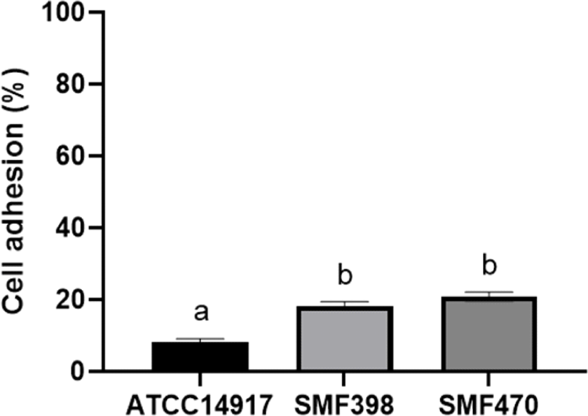
유산균이 체내에서 지속적으로 유용한 효과를 발휘하기 위해서는 장 세포에 대한 부착 능력이 요구된다(Jeong et al., 2021). 인간 결장 유래 HT-29 세포에 대한 유산균의 장 부착능은 Fig. 1과 같다. SMF398과 SMF470 균주의 HT-29 세포에 대한 부착능은 각각 17.81%와 20.71%로 ATCC14917(8.20%)과 비교하여 유의적으로 높은 장 부착능을 보였다(p<0.05). 발효식품 유래 프로바이오틱스 균주인 L. plantarum Ln1의 HT-29 세포에 대한 장 부착능은 2.69%(Jang et al., 2018), L. plantarum BSM-2와 L. plantarum EHJ-1의 HT-29 세포에 대한 장 부착능은 각각 4.01%, 4.62%로 보고된 바 있어(Jung and Kim, 2015), 본 연구에서 선별된 SMF398과 SMF470의 장 부착력은 우수하다고 볼 수 있다. Probiotic 유산균은 장 점막세포 또는 미세융모에 부착하여 군락을 형성하고, mucin의 생성을 촉진시키며, 장 투과성을 감소시키는 작용을 함으로써 병원성 미생물의 부착 및 유해 물질의 투과를 억제한다고 알려졌다(Saulnier et al., 2009).

labp-8-2-66-g1
Fig. 1. Adhesion ability of LAB strains to human intestinal epithelial cells (HT-29). Each value represents mean±standard deviation of three independent experiments. a-b Different letters indicate significant difference (p<0.05). ATCC14917; L. plantarum ATCC14917, SMF398; L. plantarum SMF398, SMF470; L. plantarum SMF470
Download Original Figure
유산균의 항균활성 및 co-aggregation 능력

선별된 유산균의 병원성 지시세균에 대한 항균활성 및 co-aggregation 능력을 측정한 결과는 Table 3과 같다. E. coli KCTC1039에 대한 항균활성 측정 결과, SMF398과 SMF470은 ATCC14917(12 mm)과 유사한 11 mm의 저해환을 생성하였다. S. aureus KCCM11335에 대하여 ATCC14917은 5 mm, SMF470는 7.67 mm의 저해환을 보인 반면, SMF398은 12 mm의 가장 높은 저해환을 생성하였다. 유산균은 대사산물로 젖산과 아세트산 등의 유기산, 박테리오신, 과산화수소 등의 항균물질을 생성하여 병원성균의 생육을 억제한다고 알려져 있다(Yuk et al, 2019).

Table 3. Antimicrobial activity and co-aggregation of LAB strains to E. coli and S. aureus
Indicator strains L. plantarum ATCC14917 L. plantarum SMF398 L. plantarum SMF470
Inhibition zone (mm) E. coli KCTC1039 12.00±0.00b 11.00±0.00a 11.00±0.00a
S. aureus KCCM11335 5.00±0.00a 12.00±0.00c 7.67±0.58b
Co-aggregation (%) E. coli KCTC1039 30.40±0.52a 49.02±0.51c 44.85±0.29b
S. aureus KCCM11335 33.22±0.38a 48.05±1.06b 50.45±0.16c

Each value represents mean±standard deviation of three independent experiments.

Values with different superscript letters in the same row are significantly different (p<0.05).

Download Excel Table

SMF398과 SMF470은 E. coli KCTC1039와 S. aureus KCCM11335에 대하여 높은 co-aggregation 능력을 나타내는 것으로 확인되었다. SMF398과 SMF470는 E. coli에 대하여 각각 49.02%와 44.85%, S. aureus KCCM11335에 대하여 각각 48.05%와 50.45%의 co-aggregation 능력을 보였으며, 두 균주 모두 ATCC14917(30.4%, 33.22%)과 비교하여 유의적으로 높은 상호응집력을 나타내었다(p<0.05). Kang 등(2017)은 발효식품에서 분리한 Lacticaseibacillus paracasei MG310이 E. coli KCCM41300에 대하여 11 mm의 생육저해환을 나타내었다고 보고하였고, Tareb 등(2013)Lacticaseibacillus rhamnosus CNCM-I-3698과 Companilactobacillus farciminis CNCM-I-3699가 E. coli K88에 대해 최대 38.2%의 co-aggregation 능력을 나타냈다고 보고한 바 있다. 따라서 본 연구에서 선별된 SMF398과 SMF470 균주는 병원균에 대한 항균활성 및 상호응집력이 우수한 것으로 판단된다.

유산균 사균체의 세포독성

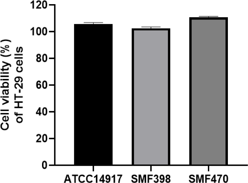
MTT assay는 살아있는 세포에 MTT tetrazolium 시약을 처리하면 세포의 mitochondria reductase에 의해 tetrazolium가 환원되어 청자색의 MTT formazan 결정을 생성하는 원리이다(Liu et al., 1997). MTT assay를 이용하여 인간 결장 유래 HT-29 세포에 각 유산균의 열처리 사균체를 1 mg/mL(약 1×10⁹ CFU/mL)의 농도로 처리한 후 HT-29 세포의 생존율을 평가한 결과, 모든 사균체 시료에 대하여 100% 이상의 생존율을 보였다(Fig. 2). 따라서 본 연구에서는 장 세포에 대하여 세포독성을 나타내지 않는 1 mg/mL 농도의 열처리 사균체를 이용하여 항산화 및 항염 활성을 평가하였다.

labp-8-2-66-g2
Fig. 2. Effect of heat-killed LAB on the viability of HT-29 cells. Heat-killed cells (1 mg/mL) of LAB were prepared by heating at 121°C for 15 min. Each value represents mean±standard deviation of three independent experiments. ATCC14917; L. plantarum ATCC14917, SMF398; L. plantarum SMF398, SMF470; L. plantarum SMF470.
Download Original Figure
유산균 사균체의 항산화 활성

유산균 사균체의 항산화 활성은 DPPH 라디칼 소거능, ABTS 라디칼 소거능 및 β-carotene bleaching 억제능을 측정하여 평가하였다(Fig. 3). DPPH 라디칼과 ABTS 라디칼은 비교적 안정한 자유라디칼로, DPPH 라디칼 소거능과 ABTS 라디칼 소거능이 높다면 자유라디칼을 환원시키거나 소거시키는 항산화 활성이 높다는 것을 의미한다(Lee and Kang, 2020). SMF398과 SMF470 사균체의 DPPH 라디칼 소거능은 각각 19.25%와 21.88%로 ATCC14917 사균체(17.8%)보다 유의적으로 높은 소거능을 나타내었다(p<0.05). ABTS 라디칼 소거능의 경우에도 SMF398 사균체는 46.16%, SMF470 사균체는 36.28%로 ATCC14917 사균체(24.82%)보다 유의적으로 높은 소거능을 보였다(p<0.05). β-Carotene bleaching assay는 리놀레산이 산화되어 생기는 지질 라디칼이 염색제인 β-carotene의 이중결합을 파괴하여 탈색을 유발하는 원리이다(Jung et al., 2016). SMF398과 SMF470 사균체의 β-carotene bleaching 억제능은 각각 43.19%와 42.88%로 ATCC14917 사균체(22.73%)보다 유의적으로 높은 지질과산화 억제능을 나타내었다(p<0.05).

labp-8-2-66-g3
Fig. 3. Effect of heat-killed LAB on DPPH radical scavenging activity (A), ABTS radical scavenging activity (B), and β-carotene bleaching inhibitory activity (C). Heat-killed cells (1 mg/mL) of LAB were prepared by heating at 121°C for 15 min. Each value represents mean±standard deviation of three independent experiments. a-c Different letters indicate significant difference (p<0.05). ATCC14917; L. plantarum ATCC14917, SMF398; L. plantarum SMF398, SMF470; L. plantarum SMF470.
Download Original Figure

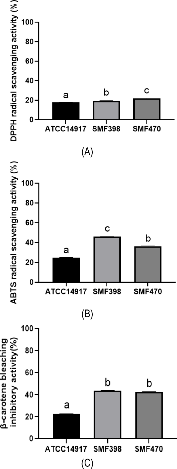
Liu와 Pan(2010)은 8종의 Lactobacillus 사균체가 14.81-28.27% 범위의 DPPH 라디칼 소거능을 나타내었고, Lee와 Kang(2020)은 8종의 Lactobacillus 사균체가 최대 28.71%의 ABTS 라디칼 소거능을 나타내었다고 보고하였다. Jang 등(2018)L. plantarum Ln1 사균체가 58.33%의 지질과산화 억제능을 나타내었다고 보고한 바 있다. 따라서 본 연구 결과, 선별된 SMF398과 SMF470 사균체는 항산화활성이 우수하며 향후 기능성 항산화제로 활용 가능할 것으로 기대된다. 유산균의 항산화 활성은 균체 표면에 존재하는 EPS 물질, 세포질의 polysaccharide, 대사물질로 생성된 glutathione과 α-tocopherol 등이 관여하며(Kim and Park, 2021), 열처리에 의해 항산화 활성을 나타내는 Maillard 반응물이 생성된다고 알려졌다(Lee and Kang, 2020).

유산균 사균체의 NO 생성 억제

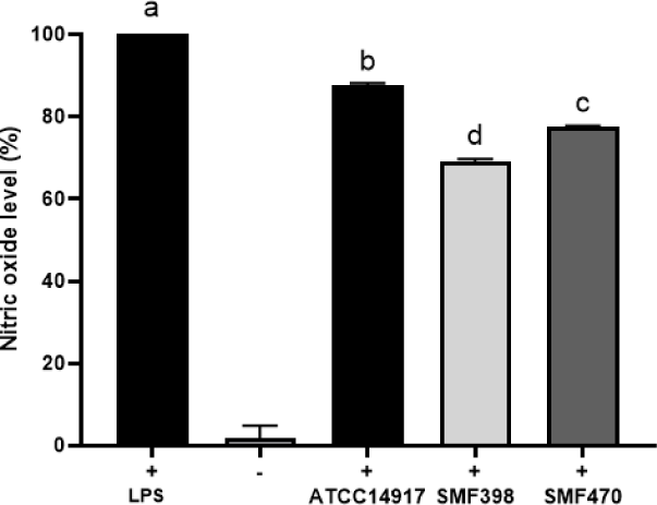
LPS 등 외부자극에 의해 과도하게 활성화된 대식세포는 L-arginine으로부터 nitric oxide(NO)의 과도한 생성을 촉진하여 염증을 악화시키고 조직을 손상시키며 병적 반응을 일으킨다(Kim et al., 2009). 유산균 사균체의 NO 생성 억제능을 평가하기 위하여, LPS 처리에 의해 염증이 유도된 RAW264.7 대식세포에 유산균 사균체(1 mg/mL)를 첨가한 후 NO 생성량을 분석하였다(Fig. 4). LPS 비처리군(-)과 비교하여 LPS 처리 대조군(+)에서 현저하게 높은 NO 생성량이 관찰되어 LPS에 의해 염증이 유도된 것이 확인되었다. SMF398과 SMF470 사균체 처리에 의해 LPS로 자극된 RAW 264.7 세포에서 NO 생성이 각각 30.92%와 22.81% 감소되었으며, ATCC14917 사균체(12.5%)보다 유의적으로 높은 NO 생성 억제능을 나타내었다(p<0.05). Son과 Kang(2021)은 물김치에서 분리한 L. plantarum P100 균주의 열처리 사균체가 22.32%의 NO 생성 억제능을 나타내었다고 보고한 바 있다.

labp-8-2-66-g4
Fig. 4. Effect of heat-killed LAB on production of nitric oxide in LPS-stimulated RAW264.7 cells. Heat-killed cells (1 mg/mL) of LAB were prepared by heating at 121°C for 15 min. Each value represents mean±standard deviation of three independent experiments. a-d Different letters indicate significant difference (p<0.05). +; LPS(1 μg/mL), -; without LPS, ATCC14917; L. plantarum ATCC14917, SMF398; L. plantarum SMF398, SMF470; L. plantarum SMF470.
Download Original Figure
유산균 사균체의 TNF-α, IL-6, iNOS, COX-2 유전자 발현 억제

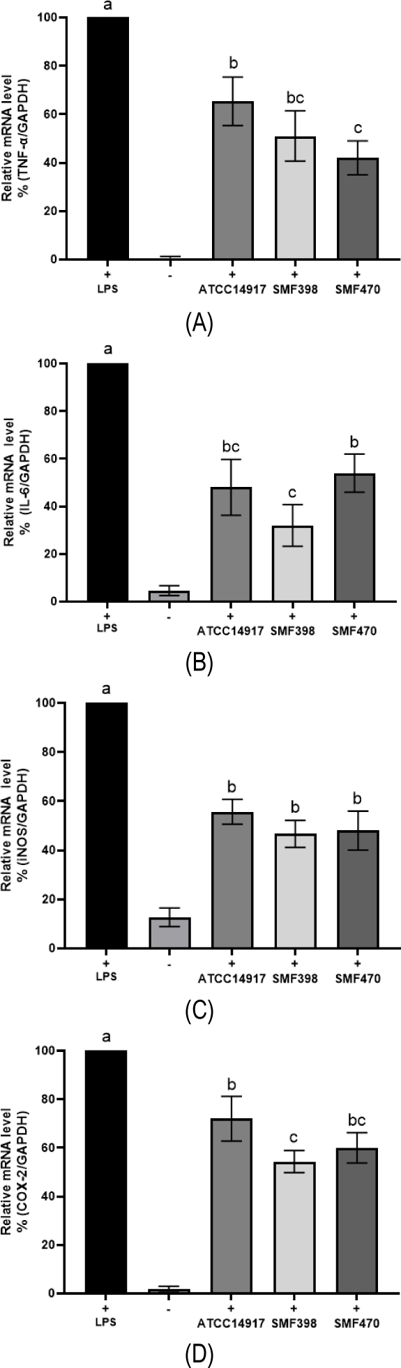
LPS와 같은 면역자극물질 또는 과량의 산화적 스트레스에 의해 대식세포가 과도하게 활성화 될 경우, tumor necrosis factor-α(TNF-α), interleukin-6(IL-6), inducible nitric oxide synthase (iNOS), cyclooxygenase-2(COX-2) 등의 염증성 매개인자들의 과도한 발현을 촉진하여 염증을 악화시키고 만성염증을 유발하여 각종 질환의 발병 요인으로 작용한다(Lawrence et al., 2002). 따라서 염증성 매개인자의 발현 억제는 염증성 질환의 예방과 치료를 위한 주요 목표가 된다. TNF-α와 IL-6는 염증성 cytokine으로 이들의 과도한 발현은 염증반응을 유발한다고 알져졌다(Cui et al., 2020; Ishihara and Hirano, 2002). NO는 염증반응의 대표적인 지표물질로 주로 iNOS의 효소작용에 의해 L-arginine으로부터 합성되며, 과도하게 생성된 NO는 염증 유발, 조직 손상, 신경 손상 등을 유발할 수 있다(Imene et al, 2016). COX-2는 iNOS와 더불어 염증반응에 주요 역할을 하는 효소로 주로 염증 부위에서 발현되어 arachidonic acid로부터 통증과 발열에 관여하는 prostaglandin E2의 합성을 유도한다(Hawkey, 1999).

유산균 사균체의 항염 활성을 평가하기 위하여, LPS 처리에 의해 염증이 유도된 RAW264.7 대식세포에 사균체를 첨가한 후 TNF-α, IL-6, iNOS, COX-2의 발현 변화를 qRT-PCR을 이용하여 측정하였다(Fig. 5). 세포 환경의 여러 조건에서도 발현 정도에 차이가 거의 없는 house keeping gene인 GAPDH를 reference gene으로 사용하였다. LPS에 의해 RAW264.7 세포에서 TNF-α, IL-6, iNOS, COX-2의 유전자 발현이 현저하게 증가하였으나, 사균체 첨가에 의해 그 발현도가 유의적으로 억제되었다(p<0.05). LPS 단독처리군과 비교하여, LPS+SMF398 사균체 또는 LPS+ SMF470 사균체 첨가군에서 TNF-α, IL-6, iNOS, COX-2의 발현이 각각 최대 58.02%, 67.70%, 53.44%, 45.90%, 저해되는 것으로 나타났다. 반면, LPS+ATCC14917 사균체 첨가군의 경우 TNF-α, IL-6, iNOS, COX-2의 발현이 각각 34.86%, 51.67%, 44.36%, 28.06% 억제되었다. 따라서 본 연구에서 선별한 유산균의 열처리 사균체는 염증성 매개인자들의 발현 억제 활성이 우수하다고 판단된다. Kang 등(2021)L. plantarum MG5203 사균체가 5×10⁸ CFU/mL의 농도에서 약 20%의 iNOS 발현 억제와 60%의 COX-2 발현 억제를 나타내었다고 보고하였고, Chon 등(2009)L. plantarum KFCC11389P 사균체가 5×10⁶ CFU/mL의 농도에서 74%의 IL-6 발현 억제를 나타내었고 보고하였다.

labp-8-2-66-g5
Fig. 5. Effect of heat-killed LAB on gene expression of TNF-α (A), IL-6 (B), iNOS (C) and COX-2 (D) in LPS-stimulated RAW264.7 cells. Heat-killed cells (1 mg/mL) of LAB were prepared by heating at 121°C for 15 min. Expressed mRNA levels of (A) TNF-α, (B) IL-6, (C) iNOS, (D) COX-2 was determined with (A-D) relative quantification by normalization with GAPDH. Each value represents mean±standard deviation of three independent experiments. a-c Different letters indicate significant difference (p<0.05). +; LPS(1 μg/mL), -; without LPS, ATCC14917; L. plantarum ATCC14917, SMF398; L. plantarum SMF398, SMF470; L. plantarum SMF470.
Download Original Figure

본 연구 결과, 김치에서 분리된 L. plantarum SMF398과 L. plantarum SMF470 균주는 내산성, 내담즙성, 항균활성 등 프로바이오틱스 균주 특성을 나타내었다. 특히 SMF398과 SMF470 균주의 열처리 사균체는 항산화 및 항염 활성이 우수하여 안전하게 섭취할 수 있는 천연 항산화제 및 항염제로의 개발이 가능할 것으로 기대된다.

요 약

김치로부터 유산균을 분리하고 프로바이오틱스 특성이 우수한 2종의 균주를 선별하여 열처리 사균체를 제조하고 항산화 및 항염 활성을 평가하였다. 선별된 L. plantarum SMF398과 L. plantarum SMF470 균주는 위산과 담즙염에 대한 저항성, 장세포(HT-29)에 대한 부착능, 병원성 세균에 대한 항균활성 및 공동응집력이 우수한 것으로 나타났다. SMF398과 SMF470 균주의 열처리 사균체는 1 mg/mL(약 1×10⁹ CFU/mL)의 농도에서 장세포에 독성을 나타내지 않아 안전성을 확인하였다. SMF398과 SMF470 사균체는 프로바이오틱스 유산균인 L. plantarum ATCC14917의 사균체와 비교하여 유의적으로 높은 DPPH 라디칼 소거능, ABTS 라디칼 소거능 및 β-carotene bleaching 억제능을 나타내었다(p<0.05). SMF398과 SMF470 사균체 처리에 의해 LPS로 자극된 RAW 264.7 세포에서 NO 생성이 각각 30.92%와 22.81% 감소되었으며, 염증성 매개인자인 TNF-α, IL-6, iNOS, COX-2의 발현은 각각 최대 58.02%, 67.70%, 53.44%, 45.90% 감소되었다. 따라서 본 연구 결과, 프로바이오틱스 특성을 갖는 L. plantarum SMF398과 L. plantarum SMF470 균주의 열처리 사균체를 활용하여 안전하게 섭취할 수 있는 항산화제 및 항염제로의 개발이 가능할 것으로 사료된다.

감사의 글

본 연구는 한국연구재단 기본연구지원사업(2021R1F1A1056371)의 지원을 받아 수행되었습니다.

References

1.

Ahmed A, Fanokh A, and Mahood M (2019) Phytochemical identification and anti-oxidant study of essential oil constituents of Ocimum basilicum L. growing in Iraq. Pharmacogn J.11, 724-729.

2.

Akter S, Park JH, and Jung HK (2020) Potential health-promoting benefits of paraprobiotics, inactivated probiotic cells. J. Microbiol. Biotechnol.30,477-481.

3.

Alberto A, Mattia di N, Anna P, Stefano R, Maddalena R, and Alessandra B (2012) Antioxidant properties of potentially probiotic bacteria: In vitro and in vivo activities. Applied Microbiology and Biotechnology. 97, 809-817.

4.

Almudena GR, Dolores GL, Adelaida EF, Teresa R, Begona BM, and Victoria MA (2014) Assessment of probiotic properties in lactic acid bacteria isolated from wine. Food Microbiology. 44, 200-225.

5.

Barros C, Pires R, Guimaraes J, Abud Y, Almada C, Pimentel T, Anna C, De Melo L, Duarte M, Silva M, Sant’Ana A, Freitas M, and Cruz A (2021) Ohmic heating as a method of obtaining paraprobiotics: Impacts on cell structure and viability by flow cytometry. Food Research International. 140, 110061.

6.

Cheon MJ, Lim SM, Lee NK, and Paik HD (2020) Probiotic properties and neuroprotective effects of Lactobacillus buchneri KU200793 isolated from Korean fermented foods. Int. J. Mol. Sci. 21, 1227.

7.

Choi HJ (2019) Recent advances on next-generation probiotics linked to the gut microbiom. Food Science and Industry. 52, 261-271.

8.

Choi MS, Chang SJ, Chae Y, Lee MH, Kim WJ, Iwasa M, Han KI, Kim JW, and Kim TJ (2018) Anti-inflammatory effect of heat-killed Enterococcus faecalis, EF-2001. J. Life Science. 28, 1361-1368.

9.

Chon H, Choi B, Lee E, Lee S, and Jeong G (2009) Immunomodulatory effects of specific bacterial components of Lactobacillus plantarum KFCC11389P on the murine macrophage cell line RAW264.7. J of Applied Microbiology. 107, 1588-1597.

10.

Chu DH and Kook MC (2019) Probiotics characterization of Lactobacillus species isolated from Kimchi. Top. Lact. Acid Bact. Probiotic. 5, 33-37.

11.

Chuah LO, Foo HL, Loh TC, Noorjaham B, Swee KY, Nur E, Raha AR, and Khatijah Y (2019) Postbiotic metabolites produced by Lactobacillus plantarum strains exert selective cytotoxicity effects on cancer cells. BMC Complementary and Alternative Medicine. 19, 114.

12.

Cui L, Wang Y, Wang H, Dong J, Li Z, Li J, Qian C, and Li J (2020) Different effects of cortisol on pro inflammatory gene expressions in LPS-, heat-killed E. coli-, or live E. coli-stimulated bovine endometrial epithelial cells. BMC Veterinary Research. 16, 1-8.

13.

Hawkey CJ (1999) COX-2 inhibitors. The Lancet. 353, 307-314.

14.

Imene S, Ryma R, Hayet R, and Chafia T (2016) Overview of cytokines and nitric oxide involvement in immuno-pathogenesis of inflammatory bowel diseases. Gastrointestional Pharmacology and Therapeutics. 7, 353-360.

15.

Ishihara K and Hirano T (2002) IL-6 in autoimmune disease and chronic inflammatory. Cytokine & Growth Factor Reviews. 13, 357-368.

16.

Jang HJ, Song MW, Lee NK, and Paik HD (2018) Antioxidant effects of live and heat-killed probiotic Lactobacillus plantarum Ln1 isolated from kimchi. J Food Sci Technol. 55, 3174-3180.

17.

Jeong SJ, Yang HJ, Ryu MS, Seo JW, and Jeong DY (2021) Characterization of Lactobacillus paracasei JSRL18-60 with probiotic properties as a starter in manufacturing fermented milk. Korean J. Food Preserv. 28, 147-159.

18.

Jung JE, Paik HD, Yoon HJ, Jang HJ, Jeewanthi RKC, Jee HS, Ki X, Lee NK, and Lee SK (2016) Physicochemical characteristics and antioxidant capacity in yogurt fortified with red ginseng extract. Korean J. Food Sci. 36, 412-420.

19.

Jung SE and Kim SH (2015) Probiotic properties of lactic acid bacteria isolated from commercial raw Makgeolli. Korean J. Food Sci.47, 44-50.

20.

Kang CH, Han SH, Kim YG, Jeong YA, and Paek NS (2017) Antibacterial activity and probiotic properties of lactic acid bacteria isolated from traditional fermented foods. Korean Society for Biotechnology and Bioengineering Journal. 32, 199-205.

21.

Kang CH, Kim JS, Kim HM, Park HM, and Paek NS (2021) Heat-killed lactic acid bacteria inhibit nitric oxide production via inducible nitric oxide synthase and cyclooxygenase-2 in RAW264.7 cells. Probiotics and Antimicrobial Proteins. 13, 1530-1538.

22.

Kim BY and Park SS (2021) The concepts and applications of postbiotics for the development of health functional food product. Lact. Acid Bact. Probiotics. 7, 14-22.

23.

Kim DH, Park SJ, Jung JY, Kim SC, and Byun SH (2009) Anti-inflammatory effects of the aqueous extract of Hwangnyeonhaedok-tang in LPS-activated macrophage cells. Korean J. Herbology. 24, 39-47.

24.

Kim SI, Kim IC, and Chang HC (1999) Isolation and identification of antimicrobial agent producing microorganisms and sensitive strain from soil. J. Korean Soc. Food Sci. Nutr. 28, 526-533.

25.

Kim SJ, Yoon YH, and Oh SJ (2020a) Suggestion of a safety evaluation procedure to improve probiotic safety. J. Dairy Sci.38, 99-111.

26.

Kim TY, Kim MG, Moon JS, Kwon HS, and Choi SK (2020b) Complete genome sequence of anti-inflammatory properties Bifidobacterium animalis subsp. lactis IDCC4301 isolated from infant feces. Korean Journal of Microbiology. 56, 62-64.

27.

Lawrence T, Willoughby DA, and Gilroy DW (2002) Anti-inflammatory lipid mediators and insights into the resolution of inflammation. Nature. 2, 787-795.

28.

Lee JA and Kang SM (2020) Antioxidant characteristics of Lactobacillus plantarum heat-killing acid bacteria isolated from Kkakdugi as cosmetic material. J. Kor. Soc. Cosm. 26, 913-922.

29.

Lee SH, Kwon MJ, Kang HT, Chung CW, Kim BO, and Kim JS (2015) The anti-inflammatory effects of probiotic-produced exopolysaccharide. J. Life Science. 25, 709-714.

30.

Lertcanawanichakul M, Kannai J, Wongmuang P, and Tharaporn S (2015) Cholesterol-lowering potentials of lactic acid bacteria with potential probiotic properties. J. PharmTech Research. 7, 463-470.

31.

Lim YS, Kim JY, and Kang HC (2019) Isolation and identification of lactic acid bacteria with probiotic activities from kimchi and their fermentation properties in milk. J. Milk Sci. Biotechnol. 37, 115-128.

32.

Liu CF and Pan TM (2010) In vitro effects of lactic acid bacteria on cancer cell viability and antioxidant activity. J. Food and Drug Analysis. 18, 77-86.

33.

Liu Y, Peterson D, Kimura H, and Schubert D (1997) Mechanism of cellular 3-(4,5-dimethylthiazol-2-yl)-2,5-diphenyltetrazolium bromide (MTT) reduction. J. of Neurochemistry. 69, 581-593.

34.

Moon GS (2019) Trends in studies on probiotics, prebiotics, and synbiotics. Food Science and Industry. 52, 208-219.

35.

Nataraj BH, Ali SA, Behare PV, and Yadav H (2020) Postbiotics-parabiotics: The new horizons in microbial biotherapy and functional foods. Microb. Cell. Fact.19, 168.

36.

Ou CC, Lin SL, Tsai JJ, and Lin MY (2011) Heat-killed lactic acid bacteria enhance immunomodulatory potential by skewing the immune response toward Th1 polarization. Journal of Food Science. 76, 260-267.

37.

Pique N, Berlanga M, and Miñana-Galbis D (2019) Health benefits of heat-killed (tyndallized) probiotics: an overview. Int. J. Mol. Sci. 20, 2534.

38.

Reddy VP, Zhu X, Perry G, and Smith MA (2009) Oxidative stress in diabetes and Alzheimer's disease. J. Alzheimers. Dis.16, 763-774.

39.

Roberta R, Nicoletta P, Anna P, Ananth P, and Min Y (1999) Antioxidant activity applying an improved ABTS radical cation decolorization assay. Free Radical Biology & Medicine. 26, 1231-1237.

40.

Saulnier D, Spinler J, Gibson G, and Versalovic J (2009) Mechanisms of probiosis and prebiosis: Considerations for enhanced functional foods. Current Opinion in Biotechnology. 20, 135-141.

41.

Seo YM, Kim HJ, Lee EJ, Chung CW, Sung HJ, Sohn HY, Park JY, and Kim JS (2018) Effects of pomace of Schizandra chinensis, schizandrin, and gomisin A on LPS-induced inflammatory responses in RAW264.7 cells. J. Life Science. 28, 339-344.

42.

Shida K, Kiyoshima-Shibata J, Kaji R, Nagaoka M, and Nanno M (2009) Peptidoglycan from lactobacilli inhibits interleukin-12 production by macrophages induced by Lactobacillus casei through Toll-like receptor 2-dependent and independent mechanisms. Immmunology. 128, 858-869.

43.

Shin JH, Lee JS, and Seo JG (2015) Assessment of cell adhesion, cell surface hydrophobicity, autoaggregation, and lipopolysaccharide-binding properties of live and heat-killed Lactobacillus acidophilus CBT LA1. Korean J. Microbiology. 51, 241-248.

44.

Sim JH, Oh SJ, Kim SK, and Baek YJ (1995) Comparative tests on the acid tolerance of some lactic-acid-bacteria species isolated from lactic fermented products. Korean J. Food Sci.27, 101-104.

45.

Son HR and Kang SM (2021) Evaluation of the antioxidant activity of lactic acid bacteria heat-killed cells derived from Mulkimchi. J. Kor. Soc. Cosmetol. 27, 868-879.

46.

Song MW, Jang HJ, and Paik HD (2019) Probiotic and antioxidant properties of novel Lactobacillus brevis KCCM 12203P isolated from Kimchi and evaluation of immunestimulating activities of its heat-killed cells in RAW264.7 cells. J. Microbiol. Biotechnol. 29, 1894-1903.

47.

Tareb R, Bernardeau M, Gueguen M, and Vernoux JP (2013) In vitro characterization of aggregation and adhesion properties of viable and heat-killed forms of two probiotic Lactobacillus strains and interaction with foodborne zoonotic bacteria, especially Campylobacter jejuni. J. Medical Microbiology. 62, 637-649.

48.

Wang S, Ahmadi S, Nagpal R, Jain S, Mishra S, Kavanagh K, Zhu X, Wang Z, McClain D, Kritchevsky S, Kitzman D, and Yadav H (2019) Lipoteichoic acid from the cell wall of a heat killed Lactobacillus paracasei D3-5 ameliorates aging-related leaky gut, inflammation and improves physical and cognitive functions: from C. elegans to mice. GeroScience. 42, 333-352.

49.

Wang Y, Zhou J, Xia X, Zhao Y, and Shao W (2016) Probiotic potential of Lactobacillus paracasei FM-LP-4 isolated from Xinjiang camel milk yoghurt. International Dairy Journal. 62, 28-34.

50.

Yang YL and Kim YJ (2001) Immunostimulationg exopolysaccharide with anticancer activity from Enterobacter sp. SSYL(KCTC0687BP) screened from Ulmus parvilolia. Korean J. Biotechnol. Bioeng. 16, 554-561.

51.

Yu HS, Lee NK, Choi AJ, Choe JS, Bae CH, and Paik HD (2019) Anti-inflammatory potential of probiotic strain Weissella cibaria JW15 isolated from kimchi through regulation of NF-κB and MAPKs pathways in LPS-induced RAW264.7 cells. J. Microbiol. Biotechnol. 29, 1022-1032.

52.

Yuk YS, Lee YK, and Kim GY (2019) Antagonistic inhibitory effects of probiotics against pathogenic microorganisms in vitro. J. of the Korea Academia-industrial. 20, 110-116.