Current Topic in Lactic Acid Bacteria and Probiotics
Korean Society for Lactic Acid Bacteria and Probiotics
Research Article

김치 유래 Lactiplantibacillus plantarum THK-j112의 Streptococcus mutans에 대한 항균효과 및 항생물막 효과

유두나1https://orcid.org/0000-0001-9107-9137, 이은지2https://orcid.org/0000-0002-1346-415X, 정제용1https://orcid.org/0000-0003-1358-9718, 이태후1https://orcid.org/0000-0001-9369-6542, 국무창3,*https://orcid.org/0000-0003-4098-8298
Du-Na Yu1https://orcid.org/0000-0001-9107-9137, Eun-Ji Yi2https://orcid.org/0000-0002-1346-415X, Je-Yong Jung1https://orcid.org/0000-0003-1358-9718, Tae-Hoo Yi1https://orcid.org/0000-0001-9369-6542, Moochang Kook3,*https://orcid.org/0000-0003-4098-8298
1경희대학교 생명공학원
2경기대학교 대학원 대체의학과
3배화여자대학교 식품영양학과
1Department of Biotechnology, Kyunghee University, Gyeonggi 17104, Republic of Korea
2Department of Alternative Medicine, Kyonggi University, Seoul 03746, Republic of Korea
3Department of Food & Nutrition, Baewha Women’s University, Seoul 03039, Republic of Korea
*Corresponding author : Moochang Kook, Department of Food & Nutrition, Baewha Women’s University, 34, Pirundae-ro 1-gil, Jongno-gu, Seoul 03039, Korea. Tel: +82-2-399-0765, Fax: +82-2-737-6711. E-mail: bmse153@gmail.com

Copyright © Korean Society for Lactic Acid Bacteria and Probiotics. This is an Open-Access article distributed under the terms of the Creative Commons Attribution Non-Commercial License (http://creativecommons.org/licenses/by-nc/4.0/) which permits unrestricted non-commercial use, distribution, and reproduction in any medium, provided the original work is properly cited.

Received: May 17, 2023; Accepted: Jun 09, 2023

Published Online: Jun 30, 2023

Abstract

Dental caries is an infectious disease accompanied by the destruction of teeth. It is a multifactorial disease caused by the interaction between microbes in dental plaque and food, with cariogenic Streptococcusmutans being one of the primary causes. In this study, the antimicrobial and antibiofilm activities of Lactiplantibacillus plantarum THK-j112 isolated from Kimchi, a traditional fermented food, against S. mutans were investigated. Furthermore, this paper also showed the possibility of developing this strain. After analyzing the 16S rRNA sequence, strain THK-j112 was named L. plantarum THK-j112 as it shared 99.93% similarity with L. plantarum ATCC 14917T. Both L. plantarum THK-j112 and L. plantarum ATCC 14917T demonstrated MIC values of 5 mg/mL and MBC values of 10 mg/mL against S. mutans KACC 16833T. The study confirmed that L. plantarum THK-j112 exhibited significant concentration-dependent inhibitory effects on biofilm formation, acid production, and glucosyltransferases(GTFs) production. Regarding hemolytic activity, Bacillus cereus ATCC 14579T, used as a positive control, was confirmed to cause hemolysis, whereas L. plantarum THK-j112 did not cause this phenomenon. Additionally, except for gentamicin, the antibiotic resistance test revealed a lower value than the MIC cut-off value for each antibiotic, indicating no resistance. Based on the findings, L. plantarum THK-j112 is expected to be useful in the food industry, such as functional food, as a lactic acid bacterium with antimicrobial and antibiofilm effects.

Keywords: antimicrobial; antibiofilm; Kimchi Streptococcus mutans; Lactiplantibacillus plantarum THK-j112

서 론

Streptococcus mutans는 구강 생물막(biofilm) 형성의 첫 단계에 관여하여 생물막을 발달시키는 역할을 하며, 당 대사를 통해 젖산을 생성하여 pH를 낮추고 칼슘을 유리시키는 방식으로 치아 우식을 일으키는 감염성 구강 질환의 대표 원인균으로 잘 알려져 있다(Hahn et al., 1991; Kim et al., 2020a). 특히 치아 범랑질에 생산된 생물막은 주로 S. mutans의 glucosyl- transferase(GTFs)에 의해 당을 glucosyl 단위로 중합하여 비수용성 글루칸을 생산하여 형성된 것으로 타액과 항생물질을 비롯한 외부 물질의 침입을 어렵게 만들고, 구강 세균 간의 유전적 돌연변이를 일으킬 가능성을 높이는 것으로 알려져 있다. 또한 S. mutans를 포함한 산을 생산하는 구강미생물에 의해 생산된 유기산이 생물막 내부에 머물게 하며, 내부로 침투한 외부 물질에 의해 산이 완충되는 작용을 할 수 없게 만드는 등 치아 우식을 촉진시키는 역할을 한다(Cho, 2021; Rukayadi and Hwang, 2006).

따라서 세균에 의한 치아우식증을 예방하기 위해서는 치아우식증의 주요 원인균인 S. mutans의 생육을 방해하는 것뿐 아니라, S. mutans의 생물막 형성을 억제하여 치아 우식이 가속화 되지 않도록 관리할 필요가 있다(Krzyściak et al., 2014; Lin et al., 2021).

이와 관련하여 기존의 치료 방식은 칫솔질 및 스케일링(scaling)을 통한 물리적인 치료방법과 클로르헥시딘(chlorhexidine) 및 플루오르화물(fluoride)을 함유한 구강청결제를 처방하는 화학적 치료 방식이 우세하였으나(Baker et al., 2017), 이러한 방법은 구강 미생물 생태계의 균형을 파괴시키고, 구강 조직을 손상시키는 등 지속적인 구강 건강 유지에 어려움이 있어 이를 대체할 새로운 연구의 필요성이 대두되었다(Kim et al., 2019). 이에 따라 최근 버섯추출물을 활용한 S. mutans에 대한 항균 및 항 GTFs 연구가 진행되는 등 천연물을 이용한 연구가 진행되고 있으며(Park et al., 2011), Lactiplantibacillus plantarum CCFM 8724 배양액이 Rat의 우식증을 억제하는 효과를 연구하는 등 유산균의 대사산물을 이용한 항 우식 연구도 꾸준히 진행되고 있다(Zhang et al., 2020).

이러한 노력에도 불구하고, 치아우식증은 2020년 기준 국내 질병 별 환자수 순위 4위를 차지하는 등 치아 우식을 개선하기 위한 제품 및 의약품 개발 수요가 지속적이므로, 꾸준한 연구와 제품개발로 이를 극복하려는 노력이 필수적일 것으로 판단된다(Kim et al., 2020b).

따라서 본 연구에서는 치아우식증을 유발하는 균 S. mutans에 대한 항균 효과 및 항생물막 효과가 우수한 유산균을 김치로부터 분리 및 동정하여, 치아우식증 예방을 위한 기능성 소재로서의 개발 가능성을 검토하고자 하였다.

재료 및 방법

사용 균주

연구에 사용된 Streptococcus mutans KACC 16833T, Bacillus cereus ATCC 14579TLactiplantibacillus plantarum ATCC 14917T 균주는 국립농업과학원 농업미생물은행(Korean Agricultural Culture Collection, KACC)과 American Type Culture Collection(ATCC)에서 분양 받아 사용하였다.

김치 유래 유산균의 분리 및 동정

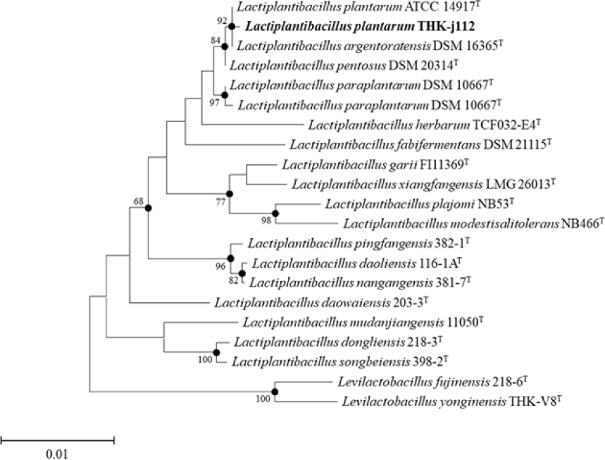
수집한 김치시료를 Lactobacilli MRS broth(Difco Co., Sparks, MD, USA) 5 mL에 1%(v/v)로 접종하여 30℃에서 24시간 종균 배양하였다. 이후 MRS broth 5 mL에 1%(v/v) 로 재 접종한 후 30℃에서 24시간 본 배양하여 유산균 감별 배지인 Bromo Cresol Purple(BCP) plate count agar(EIKEN chemical, Tokyo, Japan)에 획선 도말한 후 30℃에서 24시간 배양하였고, 산을 생성하여 colony 주위에 노란색 환을 형성한 것을 취해 순수 분리하였다. 분리한 균주의 생화학적 특성을 확인하기 위해 API 50 CHL kit(BioMérieux, Craponne, France)를 이용하여 탄수화물 발효 여부를 검토하였으며, 이후 16S rRNA 염기 서열을 분석하여 최종 동정하였다(BIOFACT Co., Daejeon, Korea). 염기서열 분석을 위해 사용한 pirmer는 27F(5'-AGAGTTTGATCMTGGCTCAG-3') 및 1492R(5'-TACGGTTACCTTGTTACGACTT-3')이며, 분석한 염기서열은 EZBioCloud website(www.ezbiocloud.net)의 16S database tool을 기반으로 표준 균주의 염기서열과 비교하여 분리 균주를 동정하였다. 계통수(phylogenetic tree)는 MEGA-X 프로그램의 neighbor-joining 및 maximum-likelihood 방법을 사용하여 작성하였다.

최소저해농도(Minimum Inhibitory Concentration, MIC)와 최소살균농도(Minimum Bactericidal Concentration, MBC) 확인

Wiegand 등(2008)의 broth dilution method를 이용하여 실험을 진행하였으며, MRS broth 5 mL에 1%(v/v) 접종하여 전배양한 유산균을 MRS broth 5 mL에 1%(v/v) 접종하여 30℃에서 24시간 본배양 하였다. 이후 4,000 rpm에서 10분간 원심분리하여 상등액을 취해 0.2 µm membrane filter(Hyundai micro Co.)로 여과한 후 농축한 것을 시료로 사용하였다. S. mutans KACC 16833T은 0.3% yeast extract를 첨가한 tryptic soy broth(TSB, Difco Co., Sparks, MD, USA)에 증균한 후 0.3% yeast extract를 첨가한 tryptic soy broth 5 mL에 1%(v/v) 접종하여 37℃에서 24시간 배양하였으며, S. mutans KACC 16833T의 세포 계수 결과를 바탕으로 0.85% NaCl을 사용하여 균수를 106cfu/mL로 조정하여 실험에 사용하였다.

96-well microplate(cell culture plate, SPL life sciences Co., Ltd, Korea)에 S. mutans KACC 16833T 배양액과 시료를 동량 분주하여 37℃에서 24시간 배양하였다. 이후 균주 배양액을 0.3% yeast extract를 첨가한 TSB agar plate에 획선 도말하여 37℃에서 24시간 배양하여 colony의 생성 여부를 확인하였다. 실험결과, colony 생성이 저해되는 농도를 최소저해농도(MIC)로 설정하였으며, colony의 생성이 확인되지 않는 농도를 최소살균농도(MBC)로 설정하였다.

생물막 형성 억제 효과

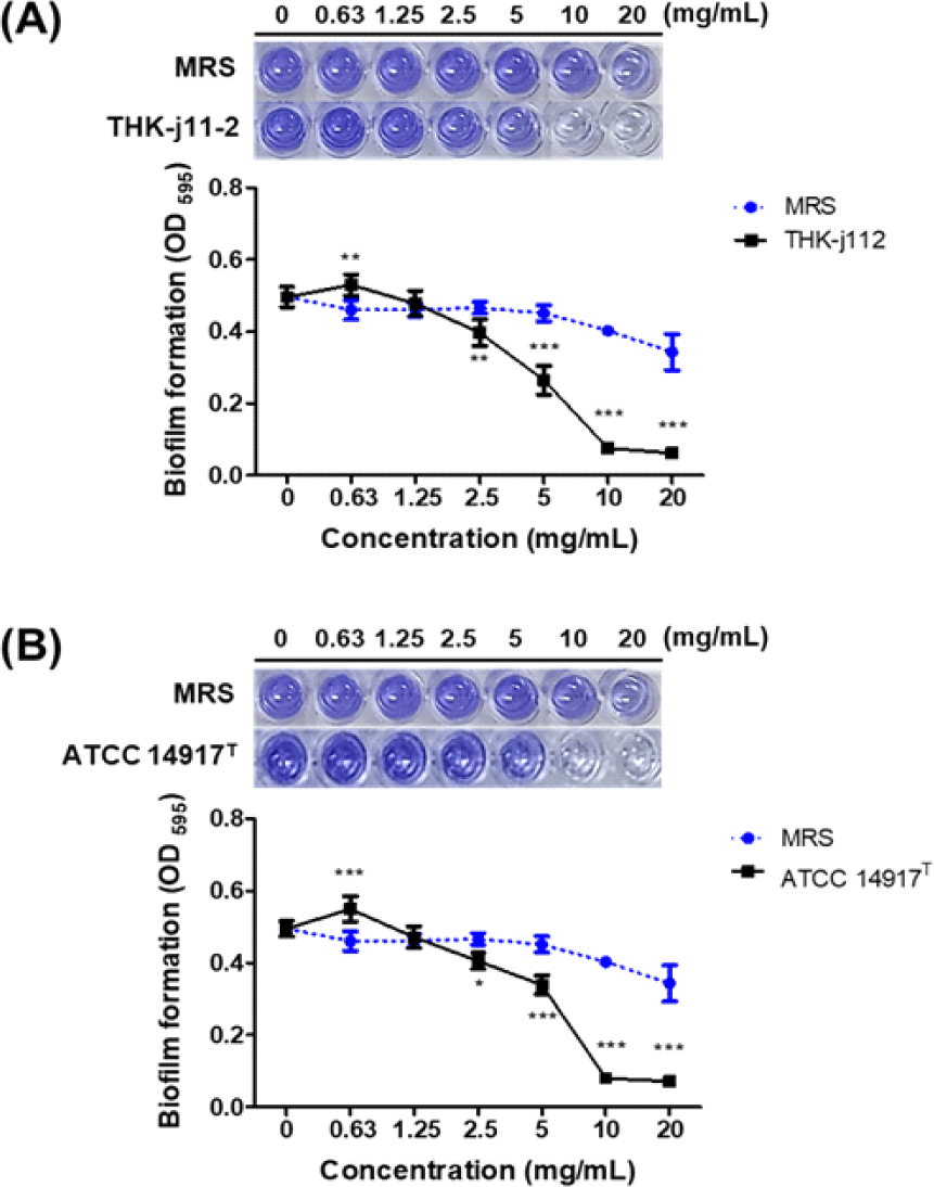
S. mutans가 생성하는 생물막 억제 효과를 확인하기 위해 Kouki et al. (2020)의 방법을 참고하여 수행하였다. S. mutans KACC 16833T을 0.3% yeast extract와 1% sucrose를 첨가한 TSB broth에 106 cfu/mL로 조정한 균액을 실험에 사용하였다. 96-well microplate에 멸균수 100 µL와 시료 100 µL를 각각 첨가하여 2-fold serial dilution한 후 균액을 100 µL분주하여 37℃에서 24시간 배양하였다. 배양 후, 배양액을 제거하고 각 well을 PBS로 2회 세척하여 건조하였으며, 0.01% crystal violet solution을 각 well에 첨가하여 15분간 염색하고 PBS로 2회 세척한 후 건조하였다. 건조된 well에 각각 33% acetic acid를 첨가하여 염색된 생물막을 용해시켜 ELISA Microplate Readers(Molecular Devices Filter Max F5; San Francisco, CA, USA)를 사용하여 595 nm에서 흡광도를 측정하였다.

유기산 억제 효과

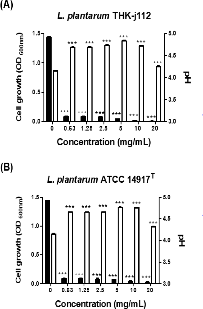
S. mutans가 생산하는 유기산은 치아에 직접적인 영향을 미쳐 치아우식을 가속화 시키는 것으로 알려져 있다. 따라서 THK-j112 균주가 S. mutans KACC 16833T의 산 생성을 억제할 수 있는지 확인하였다. 0.3% yeast extract와 1% sucrose를 첨가한 TSB broth 500 µL에 시료를 500 µL씩 첨가하여 2-fold serial dilution한 후 0.85% NaCl로 균수를 106 cfu/mL로 조정한 균액을 500 µL 분주하고 37℃에서 24시간 배양 후 pH meter(Seven Compact Duo S213; Switzerland)를 이용하여 pH를 측정하였고, UV-Vis Spectrophotometer(Mecasys Co., Seoul, Korea)를 이용하여 600 nm에서 흡광도를 측정하였다.

Glucosyltransferase(GTFs) 생성 억제 효과

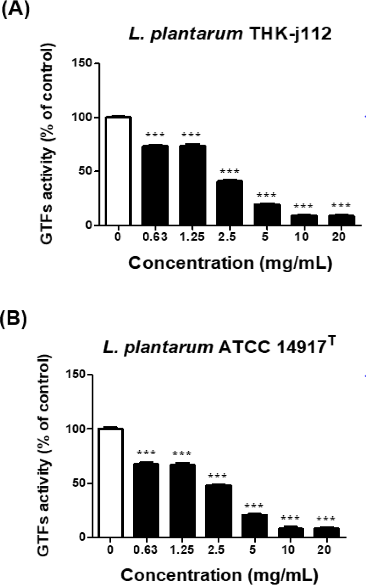
S. mutans의 세포 외 다당류를 생성하는 효소인 GTFs의 억제 정도를 확인하기 위해 GTFs의 억제 활성 평가를 실시하였으며, 실험 방법은 Mukasa et al. (1987)의 방법을 참고하였다. Sucrose를 기질로 사용하여 37℃에서 24시간 배양한 S. mutans KACC 16833T를 4,000 rpm에서 15분 동안 원심분리하고 상등액의 pH를 0.1M NaOH를 사용하여 7.0으로 보정하였다. 보정 후 0.02% sodium azide을 첨가하여 GTFs coenzyme solution을 준비하였다. 시료와 GTFs coenzyme solution 혼합액에 2.0% sucrose를 첨가하여 37℃에서 24시간 반응시킨 후 UV-Vis Spectrophotometer를 이용하여 600 nm에서 흡광도를 측정하였다.

용혈성 확인

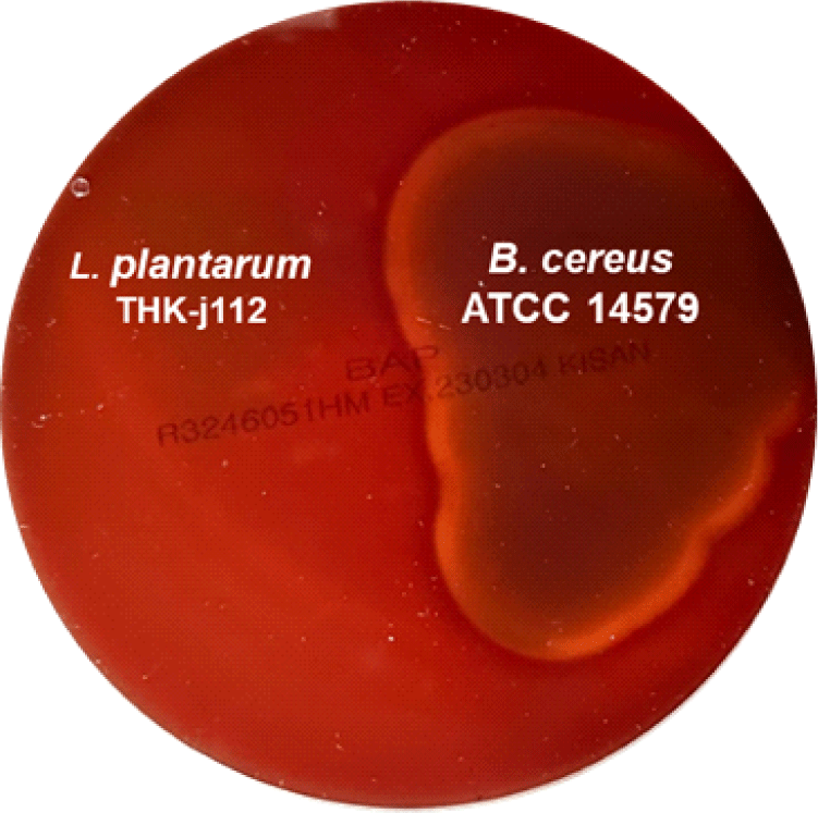
용혈성을 확인하기 위해 5% sheep blood(Kisan Bio Co., Seoul, Korea)가 함유된 blood agar plate(Kisan Bio Co., Seoul, Korea)에 분리 균주 THK-j112와 용혈성이 있는 것으로 알려진 B. cereus ATCC 14579T를 양성대조군으로 하여 획선 도말 후 37℃에서 48시간 배양하였다. 이후 colony 주위의 용혈반응을 양성대조군과 비교하여 용혈성 여부를 판단하였다.

항생제 내성 평가

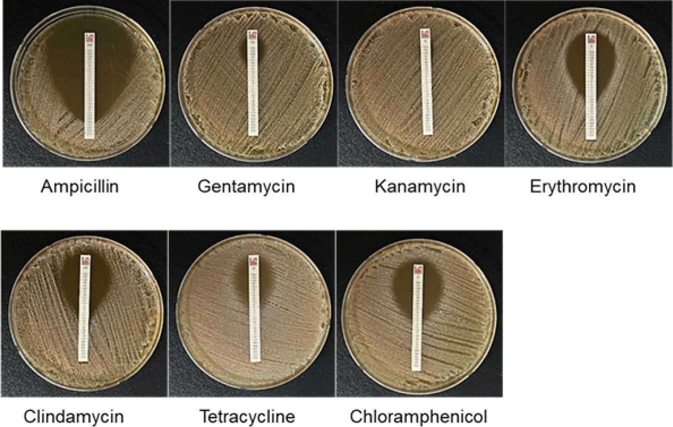
균주의 항생제 내성 특성을 파악하기 위해 유럽식품안전청(FEEDAP, 2012)의 가이드라인에 따라, E-test strip(Kisan Bio Co., Seoul, Korea)을 사용하여 MIC 값을 판독하였다. 이후 측정된 MIC 값은 EFSA 가이드라인의 cut-off value와 비교하였다.

통계분석

본 실험에서 얻은 측정치의 통계학적 분석은 Graphpad prism 5(GraphPad Software, San Diego, CA, USA)를 이용하여 평균(mean) 및 표준편차(standard deviation, SD)로 표기하였으며, 분산분석(ANOVA)을 실시하였다. Tukey의 다중비교 방법을 사용하여 사후 검증하였으며, p < 0.05 수준에서 유의성을 판단하였다.

결과 및 고찰

김치 유래 유산균의 분리 및 동정

본 실험에서 분리한 균주 THK-j112의 16S rRNA 유전자 염기서열 분석 결과, L. plantarum ATCC 14917T과 99.93%의 유사성을 나타내어 THK-j112 균주를 L. plantarum THK-j112로 명명하였다(Fig. 1). API 50 CHL kit를 이용한 L. plantarum THK-j112와 표준균주인 L. plantarum ATCC 14917T의 당 이용성 검정 결과(Table 1), 공통적인 당이용성 이외에 L. plantarum THK-j112는 L. plantarum ATCC 14917T이 이용하지 못한 D-xylose와 gentiobiose를 이용할 수 있었고, L. plantarum ATCC 14917TL. plantarum THK-j112가 이용하지 못한 mannitol, sorbitol, α-methyl-D-mannoside, melibiose, sucrose, melezitose, raffinose, D-turanose 및 D-arabitol을 이용할 수 있었다. 이러한 결과를 바탕으로, 본 실험에서 분리한 균주 THK-j112와 표준균주는 당 이용성에 대해 상당한 차이를 보이므로 생리학적, 생화학적으로 다른 특성을 가진 것으로 판단된다.

labp-9-1-9-g1
Fig. 1. Neighbor-joining phylogenetic tree of strain THK-j112. Bootstrap values(expressed as a percentage of 1,000 replications) > 65% are shown at the branch points. Bar, 0.01 substitutions per nucleotide position.
Download Original Figure
Table 1. Enzyme assay for biochemical reactions of L. plantarum THK-j112 and L. plantarum ATCC 14917T using API 50 CHL kit. Both strains are negative for acid production of glycerol, erythritol, D-arabinose, L-ribose L-xylose, adonitol D-adonitol, methyl-β-D-xylopyranosicle, L-sorbose, rhamnose, dulcitol, inositol, α-methyl-D-glucoside, inulin, starch, glycogen, xylitol, D-lyxose, D-tagatose, D-fucose, L-fucose, D-arabitol, L-arabitol, 2-keto-gluconate and 5-keto-gluconate
Substrate L. plantarum THK-j112 L. plantarum ATCC 14917T
L-Arabinose +1) +
D-Ribose + +
D-Xylose + 2)
D-Galactose + +
D-Glucose + +
D-Fructose + +
D-Mannose + +
Mannitol +
Sorbitol +
α-Methyl-D-mannoside +
N-Acethyl-glucosamine + +
Amygdalin + +
Arbutin + +
Esculin + +
Salicin + +
Cellobiose + +
Maltose + +
Lactose + +
Melibiose +
Sucrose +
Trehalose + +
Melezitose +
Raffinose +
Gentiobiose + +
D-Turanose +
Gluconate + +

+, positive,

–, negative.

Download Excel Table
MIC 및 MBC 측정

S. mutans KACC 16833T에 대하여 L. plantarum THK-j112와 L. plantarum ATCC 14917T의 최소저해농도(MIC)와 최소살균농도(MBC)를 측정하였다. 그 결과, L. plantarum THK-j112와 L. plantarum ATCC 14917T은 모두 S. mutans KACC 16833T에 대해 5 mg/mL의 MIC 및 10 mg/mL의 MBC 값을 나타내어 두 균주가 S. mutans KACC 16833T에 대해 유사한 정도의 항균활성을 보이는 것을 확인할 수 있었다.

생물막 형성 억제 효과

S. mutans KACC 16833T에 대한 L. plantarum THK-j112 및 L. plantarum ATCC 14917T의 생물막 형성 억제 효과 결과는 Fig. 2에 나타내었다. 실험결과, 두 균주 모두 배양액의 농도가 높아짐에 따라 농도 의존적으로 S. mutans KACC 16833T의 생물막 형성이 저해되어 2.5 mg/mL 이상의 농도에서 대조군 대비 유의하게 생물막 형성이 억제되는 것으로 확인되었다. 특히 5 mg/mL, 10 mg/mL 및 20 mg/mL에서 L. plantarum THK-j112의 흡광도는 0.27±0.04, 0.08±0.01 및 0.06±0.01로 확인되었으며, L. plantarum ATCC 14917T의 흡광도는 0.34±0.03, 0.08±0.00 및 0.07±0.00으로 확인되어 5 mg/mL 이상의 고농도에서 L. plantarum THK-j112와 표준균주 모두 50% 이상의 우수한 항생물막 효과를 보이는 것으로 확인되었다.

labp-9-1-9-g2
Fig. 2. Inhibition of biofilm formation of L. plantarum THK-j112 and L. plantarum ATCC 14917T. Using 1% sucrose as a substrate, the biofilm formation inhibitory effect of L. plantarum THK-j112 and L. plantarum ATCC 14917T on the biofilm produced by S. mutans was confirmed. After treatment at the indicated concentration, incubated for 24 h. The biofilm formation was measured using a microplate reader. Values are mean ± SD. * p < 0.05, ** p < 0.01 and *** p < 0.001 vs. MRS-treated group.
Download Original Figure
유기산 생성 억제 효과

S. mutans는 당 대사과정에서 유기산을 생산하여 치아 표면의 pH를 낮춰 칼슘을 용출시키는 등 치아 우식을 유발하는데 직접적인 영향을 미친다. 따라서 치아 우식을 예방하기 위해서는 S. mutans가 생산하는 유기산의 생성을 억제할 필요성이 있다. 따라서 L. plantarum THK-j112 균주가 S. mutans에 의한 산 생성을 억제하는지 여부를 pH meter와 UV-Vis Spectrophotometer를 사용하여 측정하였다. 그 결과, Fig. 3에 나타낸 바와 같이 L. plantarum THK-j112와 L. plantarum ATCC 14917T은 MBC 미만의 농도(0.63 - 5 mg/mL)에서는 농도의존적으로 산이 생성됨이 확인되었고, MBC 이상의 농도(10, 20 mg/mL)에서는 산 생성이 억제됨을 확인할 수 있었다.

labp-9-1-9-g3
Fig. 3. Inhibition of acid production of L. plantarum THK-j112 and L. plantarum ATCC 14917T. After treating S. mutans with the indicated concentration, incubated for 24 h. The amount of acid production was measured using a pH(right Y axis, □) meter and bacterial growth(left Y axis,■) was measured using a spectrophotometer. Values are mean ± SD. *** p < 0.001 vs. untreated group.
Download Original Figure
Glucosyltransferase(GTFs) 억제 효과

GTFs 억제 효과 실험결과, L. plantarum THK-j112는 0.63, 1.25, 2.5, 5, 10 및 20 mg/mL에서 각각22.42%, 26.72%, 58.95%, 80.65%, 91.24% 및 91.24%의 억제율을 보였으며, L. plantarum ATCC 14917T은 0.63, 1.25, 2.5, 5, 10 및 20 mg/mL에서 각각 33.36%, 32.42%, 52.09%, 79.28%, 91.76% 및 91.90%의 GTFs 억제를 보이는 것으로 확인되었다. 이에 따라 상기 L. plantarum THK-j112 및 L. plantarum ATCC 14917T 균주 배양액은 GTFs 생산을 억제함으로서 S. mutans의 생물막 형성을 억제할 것으로 생각되며, 특히 5 mg/mL 이상의 농도에서 L. plantarum THK-j112 및 L. plantarum ATCC 14917T 모두 90% 이상의 높은 억제율을 보여 효과적으로 S. mutans에 의한 생물막 형성 억제에 기여할 것으로 기대된다(Fig. 4).

labp-9-1-9-g4
Fig. 4. Inhibition of GTFs activity of L. plantarum THK-j112 and L. plantarum ATCC 14917T. GTFs inhibitory activity was measured using a spectrophotometer of water-insoluble glucan produced by S. mutans using sucrose as a substrate. GTFs was pretreated with the indicated concentrations for 24 h. Values are mean ± SD. *** p < 0.001 vs. untreated group.
Download Original Figure
L. plantarum THK-j112의 용혈성 확인

유산균 및 대사산물을 식품산업에 활용하기 위해서는 용혈성 및 항생제 내성과 같은 안전성 평가가 필수적이다. 따라서 본 연구에서는 L. plantarum THK-j112의 용혈성 여부를 확인하였다. 용혈이란 적혈구가 파괴되어 헤모글로빈이 혈구 밖으로 용출되는 현상으로, 혈액 한천 배지 상에서 나타나는 용혈의 유형에 따라 α, βγ-용혈로 구분할 수 있다. α-용혈은 적혈구의 부분적 용해를 말하며, 혈액배지 상에서 colony 주변이 녹색으로 변하는 특징이 있으며, β-용혈은 적혈구가 완전히 용해되는 것으로 혈액배지 상에서 colony 주변이 투명하게 변한다. γ-용혈는 용혈이 일어나지 않는 반응으로 colony 주변에 아무런 변화가 없는 것이 특징이다(Kim et al., 1995). 이러한 기준에 따라 용혈성을 확인한 결과, L. plantarum THK-j112는 용혈을 보이지 않은 반면, 양성대조군인 B. cereus ATCC 14579Tβ-용혈을 일으키는 것으로 확인되었다(Fig. 5). 따라서 L. plantarum THK-j112는 용혈을 일으키지 않는 균으로 해당 안전성 평가기준에 부합하는 것으로 확인되었다.

labp-9-1-9-g5
Fig. 5. Hemolysis of L. plantarum THK-j112 isolated from Kimchi
Download Original Figure
L. plantarum THK-j112의 항생제 내성 확인

항생제 내성 평가 결과는 Table 2Fig. 6에 나타내었다. EFSA 가이드라인에서 제시한 식품 적용 가능 미생물의 항생제 내성 기준치(cut-off value)와 비교하였을 때, Ampicillin, Kanamycin, Erythromycin, Clindamycin, Tetracycline 및 Chloramphenicol은 EFSA 가이드라인 기준치보다 MIC 값이 낮거나 같은 것으로 확인되었다. 반면, Gentamicin은 MIC 값이 기준치인 16 mg/mL 보다 높은 32 mg/mL 로 확인되었다. 따라서 향후 기능성 식품 등으로 개발하기 위해서는 L. plantarum THK-j112의 항생제 내성 유전자를 분석하고 내성 유전자의 획득성(acquired resistance) 또는 내재성(intrinsic resistance) 여부를 확인해야 할 것으로 사료된다.

Table 2. Antibiotic susceptibility values for L. plantarum THK-j112 obtained by MIC test
Antibiotics1) AMP GEN KAN ERY CLI TET CHL
MIC cut-off value(mg/mL) 2 16 64 1 2 32 8
Detection value(mg/mL) 0.47 32 48 1 1 8 4

AMP: ampicillin, GEN: gentamycin, KAN: kanamycin, ERY: erythromycin, CLI: clindamycin, TET: tetracycline, CHL: chloramphenicol

Download Excel Table
labp-9-1-9-g6
Fig. 6. Comparison of MIC test of L. plantarum THK-j112 using various antibiotics.
Download Original Figure

요약

본 연구에서는 김치에서 분리한 유산균 L. plantarum THK-j112의 치아우식증 유발균 S. mutans에 대한 항균효과, 생물막 형성 억제효과 및 안전성 평가를 수행하였다. 본 연구에서 분리한 균주 THK-j112는 치아우식균에 대해 우수한 항균력을 보였으며, S. mutans KACC 16833T에 대한 MIC는 5mg/mL, MBC는 10 mg/mL인 것으로 확인되었다. 생물막 형성 억제 실험 결과, THK-j112 배양액은 2.5 mg/mL 이상에서 대조군 대비 유의미한 억제효과를 확인할 수 있었으며, 생물막 형성을 촉진하는 역할을 하는 GTFs를 농도 의존적으로 억제하였으며, S. mutans가 생산하는 유기산을 대조군 대비 유의미하게 억제하는 것으로 확인되었다. 안전성 평가결과, THK-j112는 용혈성을 보이지 않았으나, 항생제 내성평가에서 일부 항생제에서 기준치 이상의 항생제 내성이 확인되었다. 따라서 본 연구에서 분리한 L. plantarum THK-j112는 치아우식증 유발균의 생육 및 생물막 형성을 효과적으로 억제하며, GTFs 및 유기산 생성을 억제하여 치아우식증 예방효과가 있는 기능성 제제로서 활용가능성이 있으나, 프로바이오틱스 등 식품산업에서 활용하기 위해서는 향후 내성 유전자 연구를 통해 항생제의 획득성 여부를 확인하는 추가 연구가 반드시 필요할 것으로 판단된다.

References

1.

Adams MR, and Marteau P (1995) On the safety of lactic acid bacteria from food. Int. J. Food Microbiol. 27(2-3), 263-264.

2.

Bowen WH, Burne RA, Wu H and Koo H (2018) Oral biofilms: Pathogens, matrix, and polymicrobial interactions in microenvironments. Trends Microbiol. 26(3), 229–242.

3.

Cho SJ (2021) New approaches to the control of pathogenic oral bacteria. J. Life Sci. 31(1), 100-108.

4.

Cugini C, Shanmugam M, Landge N and Ramasubbu N (2019) The role of exopolysaccharides in oral biofilms. J. Dent. Res. 98(7), 739–745.

5.

Curragh HJ and Collins MA (1992) High levels of spontaneous drug resistance in Lactobacillus. J. Appl. Bacteriol. 73(1), 31–36.

6.

Diep DB, Godager L, Brede D and Nes IF (2006) Data minig and characterization of a novel pediocin-like bacteriocin system from the genome of Pediococcus pentosaceus ATCC 25745. Microbiology. 152(6), 1649-1659.

7.

EFSA Panel on Additives Products or Substances used in Animal Feed (FEEDAP) (2012) Guidance on the assessment of bacterial susceptibility to antimicrobials of human and veterinary importance. EFSA J. 10(6), 2740.

8.

Gibbson RJ and Van Houte J (1973) On the formation dental plaque. J. Periodontol. 44(6), 347-360.

9.

Hahn CL, Falkler WA and Minah GE (1991) Microbiological studies of carious dentine from human teeth with irreversible pulpitis. Arch. Oral Biol. 36(2), 147-153.

10.

Hamada S, Ooshima T, Torii M, Imanishi H, Masuda N, Sobue S and Kotani S (1978) Dental caries induction in experimental animals by clinical strains of Streptococcus mutans isolated from Japanese children. Microbiol. Immunol. 22(6), 301-14.

11.

Kim HK, Song SO, Noh J, Jeong IK and Lee BW (2020b). Data configuration and publication trends for the Korean national health insurance and health insurance review & assessment database. Diabetes Metab. J.44(5), 671-678.

12.

Karahan AG, Kilic GB, Kart A, Aloglu HS, Oner Z, Aydemir S, Erkus O, and Harsa S (2010) Genotypic identification or some lactic acid bacteria by amplified fragment length polymorphism analysis and investigation of their potential usage as starter culture combinations in Beyaz cheese manufacture. J. Dairy Sci. 93(1), 1-11.

13.

Kim AR, Ahn KB, Yun CH, Park OJ, Perinpanayagam H, Yoo YJ, Kum KY, and Han SH (2019) Lactobacillus plantarum lipoteichoic acid inhibits oral multispecies biofilm. J. Endod. 45(3), 310-315.

14.

Kim HJ, Lee JH, Ahn DU and Paik HD (2020a) Anti-biofilm effect of egg yolk phosvitin by inhibition of biomass production and adherence activity against Streptococcus mutans. Food Sci. Anim. Resour. 40(6), 1001-1013.

15.

Kim YM, Park UK, Mok JS, and Chang DS (1995) Physiological characteristics of Listeria monocytogenes YM-7. Kor. Fish Aquatic Sci. 28(4), 443-450.

16.

Klein MI, Hwang G, Santos PH, Campanella OH and Koo H (2015) Streptococcus mutans-derived extracellular matrix in cariogenic oral biofilms. Front Cell Infect. Micrbiol. 5, 10.

17.

Koo H, Falsetta ML and Klein MI (2013) The exopolysaccharide matrix: a virulence determinant of cariogenic biofilm. J. Dent. Res. 92(12), 1065–1073.

18.

Krzyściak W, Jurczak A, Kościelniak D, Bystrowska B and Skalniak A (2014) The virulence of Streptococcus mutans and the ability to form biofilms. Eur. J. Clin. Microbiol. Infect. Dis. 33(4), 499-515.

19.

Kumar A, Alam A, Rani M, Ehtesham NZ and Hasnain SE (2017) Biofilms: survival and defense strategy for pathogens. Int. J. Med. Microbiol. 307(8), 481–489.

20.

Lin Y, Chen J, Zhou X and Li Y (2021) Inhibition of Streptococcus mutans biofilm formation by strategies targeting the metabolism of exopolysaccharides. Crit. Rev. Microbiol. 47(5), 667-677.

21.

Mathara JM, Schillinger U, Guigas C, Franz C, Kutima PM, Mbugua SK, Shin HK and Holzapfel WH (2008) Functional characteristics of Lactobacillus spp. from traditional Maasai fermented milk products in Kenya. Int. J. Food Microbiol. 126, 57-64.

22.

Miyoshi M (1987) Antieurodontic effect of various fractions extracted from the leaves of Gymnema sylvestre. J. Yonago Med. Assoc. 38, 127-137.

23.

Mosci F, Perito S, Bass S, Capuano A and Marconi PF (1990) The role of Streptococcus mutans in human caries. Minerva Stomatol. 39, 413-429.

24.

Nakamura K, O'Neill AM, Williams MR, Cau L, Nakatsuji T, Horswill AR and Gallo RL (2020) Short chain fatty acids produced by Cutibacterium acnes inhibit biofilm formation by Staphylococcus epidermidis. Sci. Rep. 10(1), 21237.

25.

Park EJ, Lee JS and Choi WS (2011) Anti-cariogenic activities of mushroom extracted with various solvent systems. Korean Journal of Food Science and Technology. 43(6), 783–786.

26.

Rukayadi Y and Hwang JK (2006) In vitro activity of xanthorrhizol against Streptococcus mutans biofilms. Lett. Appl. Microbiol. 42(4), 400-404.

27.

Saarela M, Mogensen G, Fondén R, Mättö J and Mattila-Sandholm T (2000) Probiotic bacteria: safety, functional and technological properties. J. Biotechnol. 84(3), 197-215.

28.

Salminen S, von Wright A, Morelli L, Marteau P, Brassart D, de Vos WM, Fondén R, Saxelin M, Collins K, Mogensen G, Birkeland SE, and Mattila-Sandholm T (1998) Demonstration of safety of probiotics - a review. Int. J. Food Microbiol. 44(1-2), 93-106.

29.

Tagg JR, Dajani AS and Wannamake LW (1976) Bacteriocins of gram-positive bacteria. Bacteriol. Rev. 40(3), 722- 756.

30.

Takahashi N and Nyvad B (2011) The role of bacteria in the caries process: ecological perspectives. J. Dent. Res.90(3), 294–303.

31.

Wasilewski A, Zielinska M, Storr M and Fichna J (2015) Beneficial effects of probiotics, prebiotics, synbiotics, and psychobiotics in inflammatory bowel disease. Inflamm. Bowel Dis. 21(7), 1674-1682.

32.

Wiegand I, Hilpert K and Hancock RE (2008) Agar and broth dilution methods to determine the minimal inhibitory concentration (MIC) of anti-microbial substances. Nat. Protoc. 3(2), 163-175.

33.

Zhang L, Shen Y, Qiu L, Yu F, Hu X, Wang M, Sun Y, Pan Y and Zhang K (2022) The suppression effect of SCH-79797 on Streptococcus mutans biofilm formation. J. Oral Microbiol. 14(1), 2061113.

34.

Zhang Q, Qin S, Xu X, Zhao J, Zhang H, Liu Z, and Chen W (2020) Inhibitory effect of Lactobacillus plantarum CCFM8724 towards Streptococcus mutans- and Candida albicans-induced Caries in Rats. Oxid. Med. Cell Longev.